Figures & data
Figure 1. Number of publications in PubMed about skin gene therapy.
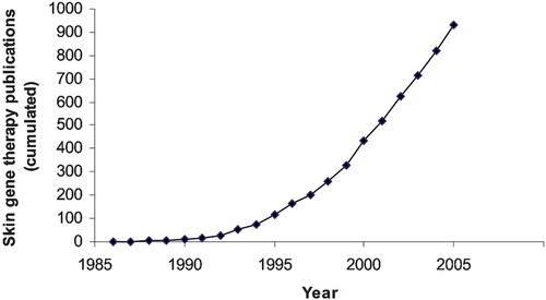
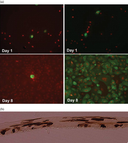
Figure 2. Combined viral and nonviral gene transfer into human keratinocytes performed as previously describedCitation50. a Monolayer cultures of human keratinocytes. The cells were transfected with a retroviral construct containing the LDL receptor gene (left panel) or cotransfected with the same construct and the pPAM3 plasmid containing gag pol and env genes (right panel). The LDL receptor protein was visualized using immunostaining at the indicated time points after transfection. (Green = FITC‐labeled LDL receptor protein; red = propidium iodine‐staining of nuclei) for colours please see (50). After cotransfection the cells produce retroviral vectors that subsequently transduce 100% of the cells. b Raft cultures of human keratinocytes cultured in the liquid‐air interphase. The cells were transfected in monolayer with a retroviral construct containing a GFP gene and the pPAM3 plasmid containing gag pol and env genes and subsequently induced to differentiate to a multilayered tissue. GFP protein was visualized using immunostaining as described Citation50. The anti‐GFP antibody was visualized using peroxidase‐staining leading to the brown color. GFP‐positive cells are found in all layers of the tissue. In control cultures transfected with the GFP construct alone no GFP‐positive cells were found at this time point (13 days after transfection) (data not shown).