Figures & data
Figure 1. ADAM-9, ADAM-15, and ADAM-17 expression in control samples from internal thoracic artery (ITA), and carotid, aortic, and femoral artery samples. Gene expression value is the normalized average gene intensity for each group measured in the Illumina Expression BeadChips. *P < 0.05, **P < 0.01, ***P < 0.001 relative to ITA, Mann-Whitney U-test.
Figure 2. ADAM-9, ADAM-15, and ADAM-17 expression in atherosclerotic samples and control samples from internal thoracic artery (ITA). Gene expression value is the normalized average gene intensity for each group measured in the Illumina Expression BeadChips. *P < 0.05, **P < 0.01, ***P < 0.001 relative to ITA, Mann-Whitney U-test.
Figure 3. Expression of ADAM-9, ADAM-15, and ADAM-17 mRNA in control samples from internal thoracic artery (ITA), and carotid, aortic, and femoral artery samples measured with TaqMan Low-Density Array. *P < 0.05, **P < 0.01, ***P < 0.001 relative to ITA, Mann-Whitney U-test.
Figure 4. Expression of ADAM-9, ADAM-15, and ADAM-17 in human atherosclerotic plaques. Serial staining of monocyte marker CD68 and smooth muscle cell marker HHF35 in human aortic (A), femoral (B), and carotid (C) plaques. Samples from internal thoracic artery (ITA) served as controls (D). NV indicates a neovascularized vessel. A straight line indicates the transition between the intima and media. Arrows indicate typical positively stained cells. The stage of atherosclerosis was classified according to American Heart Association (AHA) classification (type 1–6). 100× magnification.
Figure 5. Double immunofluorescence images demonstrating co-localization of ADAM-9 and ADAM-17 and CD68, and mirror image sections showing the co-localization of ADAM-15 and CD68. ADAM-9-immunoreactive cells at the border of adventitia and media (arrows) are also labeled with CD68. High-magnification image showing ADAM-9/CD68-immunoreactive cells (arrows) adhered to the intima of neovascularization capillary (arrowheads point to the intima exhibiting red/yellow autofluorescence). ADAM-9 labeling is very strong in the cell membrane. Mirror image sections indicating co-localization of ADAM-15 and CD68. Arrows point to the cells immunoreactive to both antigens. Double immunofluorescence photographs demonstrating the co-localization of ADAM-17 and CD68. ADAM-17 immunoreactivity is very strong in the cell membrane whereas CD68 labeling is seen throughout the cytoplasm.
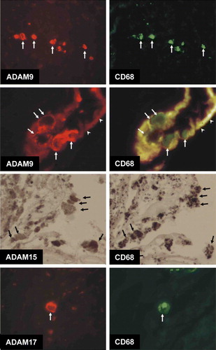
Figure 6. Western detection of (A) ADAM-9, (B) ADAM-15, and (C) ADAM-17 in atherosclerotic patient samples from carotid Citation[1], Citation[3], femoral Citation[2], and aortic Citation[4] plaques, visualized with enhanced chemiluminescence. The positions of the molecular weight markers and active/inactive forms are indicated.
![Figure 6. Western detection of (A) ADAM-9, (B) ADAM-15, and (C) ADAM-17 in atherosclerotic patient samples from carotid Citation[1], Citation[3], femoral Citation[2], and aortic Citation[4] plaques, visualized with enhanced chemiluminescence. The positions of the molecular weight markers and active/inactive forms are indicated.](/cms/asset/1cdc8f33-5b11-4774-ba61-df8f88847c1e/iann_a_365143_f0006_b.gif)