Figures & data
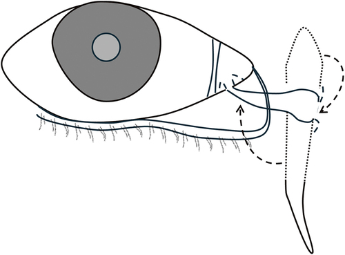
Figure 1. LT Jones’ Canaliculodacryocystostomy.
The lateral half of the fundus of the lacrimal sac was used to connect with the inferior canaliculus and therefore reconstruct the resected common canaliculus.
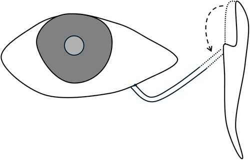
Figure 2. BR Jones’ Canaliculodacryocystorhinostomy.
Barrie Jones’ technique involved an anastomosis of the canaliculus with the lacrimal sac, which was also combined with a dacryocystorhinostomy. The dacryocystorhinostomy was used to prevent adhesions between the raw edge of the cut canaliculus and the lacrimal sac mucosa.
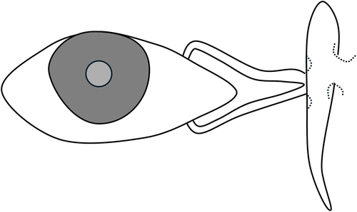
Figure 3. Stallard’s Conjunctivodacryocystostom.
The lacrimal sac was mobilized from the lacrimal fossa and brought to the plane of the caruncle. The fundus of the sac was incised and the edges sutured to the conjunctival incision edges to create a bypass from the conjunctiva to the lacrimal sac and down into the nasolacrimal duct.
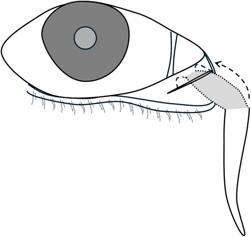
Figure 4. LT Jones’ Conjunctivodacryocystorhinostomy with Jones tube.
The original publication describes an external dacryocystorhinostomy, followed by the insertion of a polyethylene or glass tube from the medial conjunctival fornix into the nasal cavity.
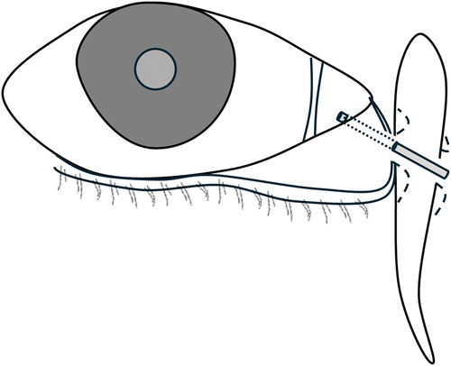
Figure 5. Conjunctivoductivodacryocystorhinostomy.
The distal end of the nasolacrimal duct is severed and the lacrimal sac is rotated approximately 90 degrees, such that the nasolacrimal duct is anastomosed with the conjunctival fornix. The residual fundus is then opened so that tears can flow from the conjunctival fornix through the neo-nasolacrimal duct, lacrimal sac and out into the nasal cavity.