Figures & data
Table 1 Distribution of findings among different methods
Figure 1. MRA shows a left renal artery that has been interpreted as stenotic.
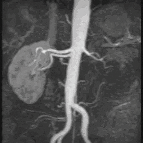
Figure 2. DSA of the same patient that demonstrates normal patency of the left renal artery.
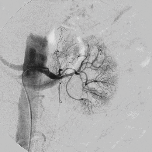
Figure 3. Transverse color Doppler US image. The peak systolic velocity is below 100cm/sec, which is considered normal.
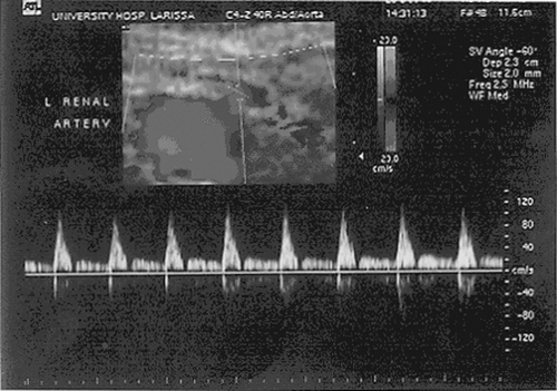
Figure 4. MRA demonstrates a severe stenosis, which was confirmed by DSA.
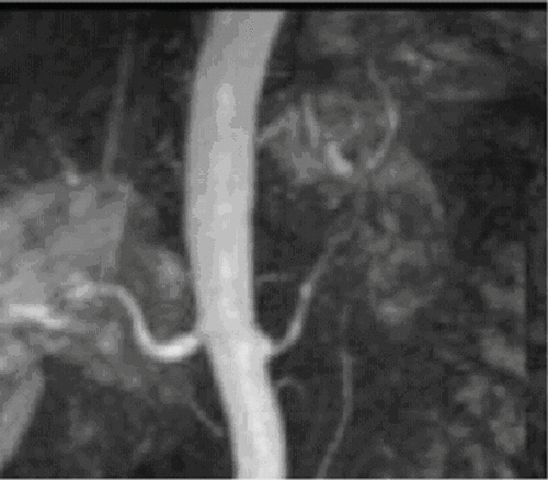
Table 2 Imaging findings of US, CTA, MRA, and DSA
Table 3 Distribution of true positive findings related to mild and severe stenosis among different methods
Table 4 Results of diagnostic efficacy of all three methodscompared with DSA