Figures & data
Figure 1. PCR analysis of the ICGN/Oa mice for the tensin2 locus. The DNA extracted from the wild-type (lane 1), heterozygous (lane 2), and homozygous (lane 3) mice were genotyped using a single pair of specific primers for the wild-type and mutant alleles, respectively. Lane M was loaded with the size marker.
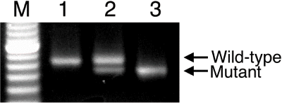
Figure 2. Effects of a tensin2 mutation on body and kidney weights of the heterozygous and homozygous mice at 5, 15, and 25 weeks of age compared with the wild-types. (A) body and (B) kidney weights. These parameters are expressed as mean ± SD. *p < 0.05, ***p < 0.001 as compared with the wild-types.
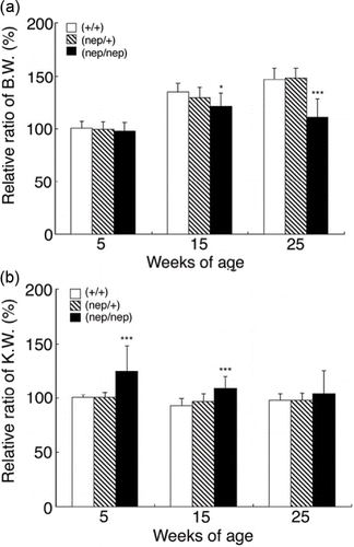
Figure 3. SDS-PAGE analysis of urine from the three strains of ICGN/Oa mice at 5, 15, and 25 weeks of age.
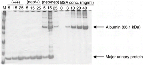
Table 1 Effects of a tensin2 mutation on urine and serum biochemical parameters in the heterozygous and homozygous mice at 5, 15, and 25 weeks of age compared with the wild-types
Figure 4. Histopathology of the renal cortex in the ICGN/Oa mice. Representative photographs of renal arteries at 5 weeks of the (A) wild-types, (B) heterozygotes, and (C) homozygotes, and at 25 weeks of the (D) wild-types, (E) heterozygotes, and (F) homozygotes. (G) Semiquantification of these histological findings. Mononuclear cell infiltration indexes are expressed as mean ± SD. *p < 0.05, ***p < 0.001 as compared with the wild-types. Representative photomicrographs of renal tubules at 5 weeks of the (H) wild-types, (I) heterozygotes, and (J) homozygotes, and at 25 weeks of the (K) wild-types, (L) heterozygotes, and (M) homozygotes. (N) Semiquantification of these histological findings. Tubular injury indexes are expressed as mean ± SD.
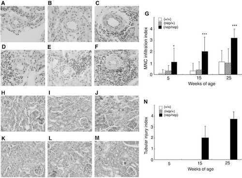
Figure 5. Time courses of the expression of α-SMA and type I collagen in the ICGN/Oa mice. Representative photomicrographs of α-SMA immunostain at 5 weeks of the (A) wild-types and (B) homozygotes, 15 weeks of the (C) wild-types and (D) homozygotes, and 25 weeks of the (E) wild-types and (F) homozygotes. (G) Semiquantification of these histological findings. Matrix scores for α‐SMA are expressed as mean ± SD. Representative photomicrographs of type I collagen immunostain at 5 weeks of the (H) wild-types and (I) homozygotes, 15 weeks of the (J) wild-types and (K) homozygotes, and 25 weeks of the (L) wild-types and (M) homozygotes. (N) Semiquantification of these histological findings. Matrix scores for type I collagen are expressed as mean ± SD. **p < 0.01, ***p < 0.001 as compared with the wild-types.
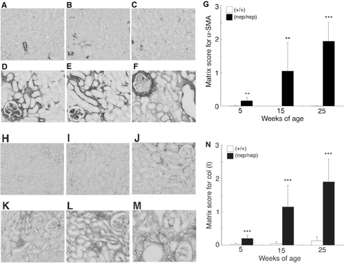
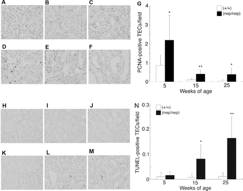
Figure 6. Time courses of the proliferating and apoptotic TECs in the ICGN/Oa mice. Representative photomicrographs of PCNA immunostain at 5 weeks of the (A) wild-types and (B) homozygotes, 15 weeks of the (C) wild-types and (D) homozygotes, and 25 weeks of the (E) wild-types and (F) homozygotes. (G) Semiquantification of these histological findings. The numbers of PCNA-positive TECs are expressed as mean ± SD. Representative photomicrographs of TUNEL at 5 weeks of the (H) wild-types and (I) homozygotes, 15 weeks of the (J) wild-types and (K) homozygotes, and 25 weeks of the (L) wild-types and (M) homozygotes. (N) Semiquantification of these histological findings. The numbers of TUNEL-positive TECs are expressed as mean ± SD. *p < 0.05, **p < 0.01 as compared with the wild-types.