
Figures & data
Figure 1. Light microscopic micrographs of rat kidney sections stained with H&E from control (A), sham (B), and experimental (D) groups. (A) Structure of kidney glomerular (star), proximal (right arrow) and distal tubule (arrowhead) with normal histological structure in the histological sections of the control group. (B) Structure of kidney glomerular (star), proximal (right arrow), and distal tubule (arrowhead) with normal histological structure in the histological sections of the sham group. (D) Atrophic glomeruli (arrowhead), tubular enlargement (star), disorganization in proximal and distal tubular epithelial cells (right arrow), tubular epithelial cell loss (left arrow).
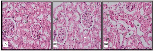
Figure 2. Light microscopic micrographs of rat kidney sections stained with PAS from control (A), sham (B), and experimental (D) rats. (A) Bowman capsule (left arrow), proximal and distal tubule basement membranes (arrowhead) and proximal tubule bristle edge structure (right arrow) of kidney glomeruli with normal histological structure in the histological sections of the control group. (B) Proximal and distal tubule basement membranes (arrowhead) and proximal tubule bristle edge structure (right arrow) with normal histological structure in the histological sections of the sham group. (D) Brush edge loss (right arrow) and dense PAS (+) mesangial cells (arrowhead) in the proximal tubule epithelium.
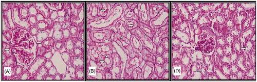
Table1. MDA and GSH levels in tissues.
Table 2. Comparison of GSH and MDA levels between groups.