Figures & data
Figure 1. Kidney under ultrasound. (A) Normal human kidney. (B) The kidney of the patient reported in our case. It showed diffuse echogenic enhancement under ultrasound, with unclear corticomedullary boundary and normal kidney size, abbreviated as ‘large white kidney’. (C) Kidney with acute ischemic injury. (D) End-stage sclerosing kidney.
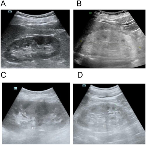
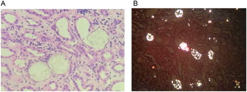
Figure 2. Pathological images of kidney biopsy. (A) In the lumen of the renal tubules, pale yellow crystalline substances are frequently seen and are considered to be oxalate crystals. The epithelial cells of the renal tubules are flattened with brush-like margins or loss of cytoplasm. The renal interstitium is infiltrated by monocytes (HE, ×10). (B) Polarized light microscopy shows polychromatic birefringence and different forms of crystals, confirming that they are oxalate crystals, widely distributed in the tubular lumen and scattered in the interstitium.