
Figures & data
Figure 1. MgH2 ameliorates APAP-induced renal dysfunction, histological injury and oxidative stress in mice.
(A, B) The levels of SCr and BUN in serum of mice were determined by corresponding kits.
(C–E) The renal histological changes in mice were observed by HE staining (C), Masson staining (D) and PAS staining (E).
(F, G) The protein expressions of NGAL, KIM-1 and iNOS in kidneys of mice were detected by western blotting.
(H) The intracellular ROS level in renal tissues of mice was detected using DHE.
The results were expressed as mean ± SEM. Statistical comparisons were performed using a Newman–Keuls test (*p < 0.05 vs. Control group, #p < 0.05 vs. APAP group).
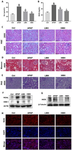
Figure 2. MgH2 improves APAP-induced inflammation and apoptosis in kidneys of mice.
(A, B) The mRNA expressions of TNF-α and IL-1β in kidneys of mice were detected by RT-qPCR.
(C, D) The protein expressions of TNF-α, IL-1β, Bad, Bax, Caspase3 and CytC in kidneys of mice were detected by western blotting.
(E) Tubular cell apoptosis in kidneys of mice was observed by TUNEL staining.
The results were expressed as mean ± SEM. Statistical comparisons were performed using a Newman–Keuls test (*p < 0.05 vs. Control group, #p < 0.05 vs. APAP group).
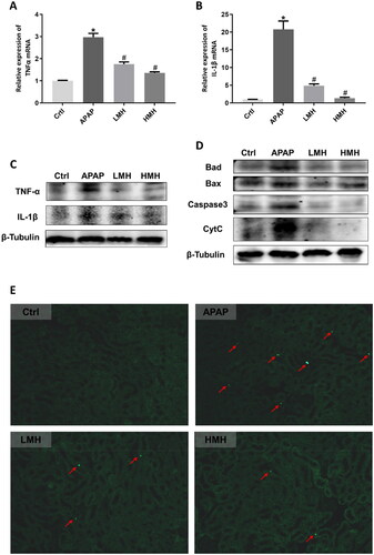
Figure 3. MgH2 alleviates APAP-induced cytotoxicity, oxidative stress, mitochondrial dysfunction and inflammation in HK-2 cells.
(A-C) HK-2 Cell viability was detected by CCK-8 assay. (A) HK-2 cells was administered with different concentrations of MgH2 (10, 20, 50, 100, 200, 400, 600, 800 and 1000 μM); (B) HK-2 cells was administered with different concentrations of APAP (5, 10, 20, 40, 60, 80, 100 mM) with or without MgH2 (0.4 mM); (C) HK-2 cells was administered with APAP (10 mM) with or without MgH2 (0.2, 0.4 mM).
(D) The ROS level in HK-2 cells was determined by DCFH-DA.
(E) The mitochondrial function of HK-2 cells was assessed by JC-1 staining.
(F, G) The protein expressions of iNOS, KIM-1 and IL-1β in HK-2 cells were detected by western blotting.
The results were expressed as mean ± SEM. Statistical comparisons were performed using t-test (*p < 0.05 vs. APAP group) or Newman–Keuls test (*p < 0.05 vs. Control group, #p < 0.05 vs. APAP group).
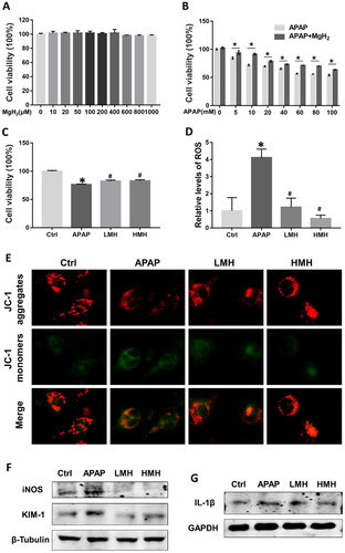
Figure 4. MgH2 suppresses APAP-induced TXNIP/NLRP3/NF-κB signaling pathway activation in HK-2 cells.
(A) The protein expressions of TXNIP, NLRP3, NF-κB p65 and p-NF-κB p65 in HK-2 cells were detected by western blotting.
(B, C) The protein expressions of TXNIP and p-NF-κB p65 in HK-2 cells were detected with immunofluorescence assay.
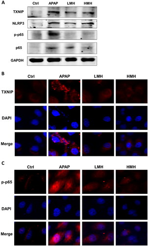
Figure 5. MgH2 suppresses APAP-induced TXNIP/NLRP3/NF-κB signaling pathway activation in kidneys of mice.
(A) The protein expressions of TXNIP, NLRP3, NF-κB p65 and p-NF-κB p65 in kidneys of mice were detected by western blotting.
(B, C) The protein expressions of TXNIP and p-NF-κB p65 in kidneys of mice were detected with immunohistochemistry staining.
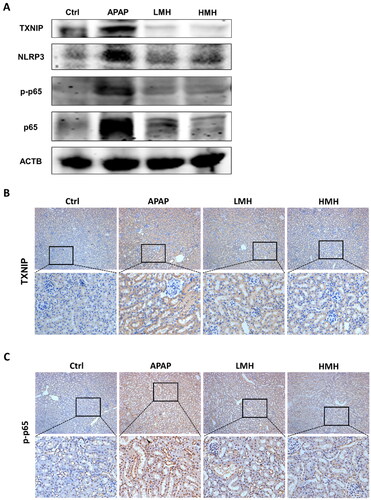
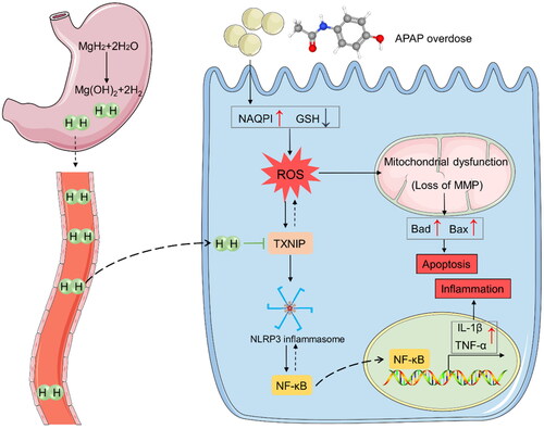
Figure 6. MgH2 protects against APAP-AKI by inhibiting TXNIP/NLRP3/NF-κB pathway. APAP overdose leads to elevated NAPQI level and GSH depletion, resulting in enhanced renal ROS production. ROS can lead to mitochondrial dysfunction and activate TXNIP/NLRP3/NF-κB signaling pathway, resulting in apoptosis and inflammation. Besides, TXNIP can further aggravate accumulation of ROS and oxidative stress. MgH2 may alleviate APAP-AKI through improving oxidative stress, inflammation and fibrosis via suppressing TXNIP/NLRP3/NF-κB pathway.
Supplemental Material
Download PDF (546.4 KB)Data availability statement
All data generated or analyzed during this study are included in this article. Further inquiries can be directed to the corresponding author.