
Figures & data
Table 1. Composition and information of SSR.
Figure 1. Construction of the 5/6 (A/I) rat model of CKD. 5/6 (A/I), 5/6 renal ablation/infarction; CKD, chronic kidney disease.
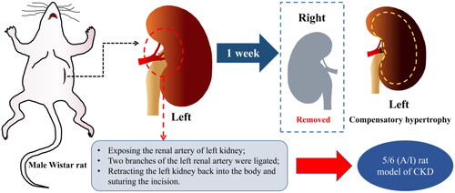
Figure 2. Flowchart of BOLD-fMRI examination in rats. BOLD-fMRI, blood oxygenation level-dependent functional magnetic resonance imaging.
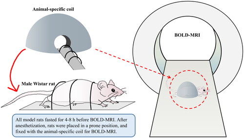
Figure 3. BOLD-fMRI T2*WI and T2* map in the coronal plane of rats. (A, B) The rat from Sham group. (C, D) The rat from 5/6 (A/I) group. (E, F) The rat from 5/6 (A/I) + LOS group. (G, H) The rat from 5/6 (A/I) + SSR group. ROI-based technique, with placement of circled ROIs in the renal cortex and medulla. BOLD-fMRI, blood oxygenation level-dependent functional magnetic resonance imaging; ROI, region of interest.
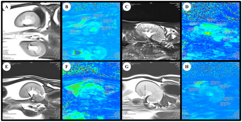
Figure 4. Bland-Altman plots of BOLD-fMRI analyses between observers. BOLD-fMRI, blood oxygenation level-dependent functional magnetic resonance imaging.
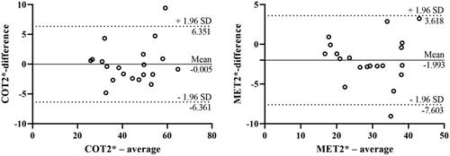
Figure 5. Effects of SSR on renal oxygenation in 5/6 (A/I) rats. (A) The level of COT2* was evaluated by BOLD-fMRI after treatment. (B) The level of MET2* was evaluated by BOLD-fMRI after treatment. (C) The expression of HIF-1α protein was determined by western blot. (D) Quantitative analysis of HIF-1α protein level (n = 4). Data were analyzed by One-way ANOVA. Values are mean ± SE. *p < 0.05; **p < 0.01; ***p < 0.001; ****p < 0.0001. COT2*, cortical T2*; MET2*, medullary T2*; LOS, Losartan; SSR, Shen Shuai II Recipe.
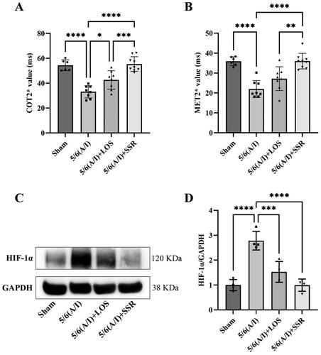
Table 2. Comparisons of COT2* and MET2* between four groups. ().
Figure 6. Effects of SSR on kidney and liver function and proteinuria in 5/6 (A/I) rats. The levels of SCr (A) and BUN (B) before treatment. The levels of SCr (C), BUN (D), CysC (E), UA (F), Ccr (G), 24-h Upr (H), UACR (I), ALT and AST (J) were measured after 8-week treatment. Data were analyzed by One-way ANOVA or Student’s t-test. Values are mean ± SE. *p < 0.05; **p < 0.01; ***p < 0.001; ****p < 0.0001. SCr, serum creatinine; BUN, blood urea nitrogen; CysC, serum cystatin C; UA, serum uric acid; Ccr, creatinine clearance rate; Upr, urinary protein; UACR, urinary albumin:creatinine ratio; ALT, alanine aminotransferase; AST, aspartate aminotransferase.
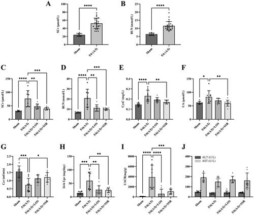
Figure 7. Effects of SSR on renal injury and fibrosis in 5/6 (A/I) rats. (A) The expression of FN, Col-I, and α-SMA proteins were determined by western blot. (B) Quantitative analysis of FN, Col-I, and α-SMA levels (n = 4). (C) Representative photomicrographs of HE and Masson’s trichrome staining. 200 × magnification. (D) The severity of tubular damage was assessed by the tubular injury score (n = 8). (E) Semiquantitative analysis of collagen area (n = 4). Data were analyzed by One-way ANOVA. Values are mean ± SE. *p < 0.05; **p < 0.01; ***p < 0.001 vs. Sham group, #p < 0.05, ##p < 0.01, ###p < 0.001 vs. 5/6 (A/I) group. LOS, Losartan; SSR, Shen Shuai II Recipe.
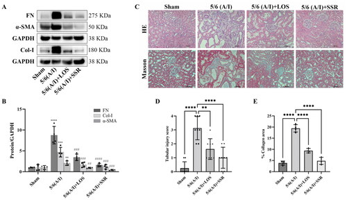
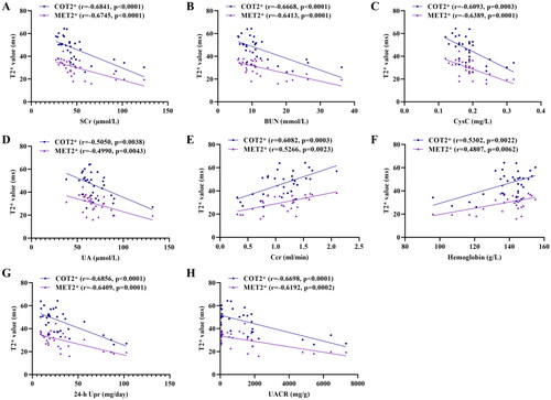
Figure 8. Correlation of COT2* and MET2* with SCr (A), BUN (B), CysC (C), UA (D), Ccr (E), Hemoglobin (F), 24-h Upr (G), and UACR (H). Data were analyzed by Pearson’s correlation coefficient. COT2*, cortical T2*; MET2*, medullary T2*; SCr, serum creatinine; BUN, blood urea nitrogen; Cys C, serum cystatin C; UA, serum uric acid; Ccr, creatinine clearance rate; Upr, urinary protein; UACR, urinary albumin:creatinine ratio.
Availability of data and materials
The datasets generated and/or analyzed during the current study are available from the corresponding author on reasonable request.