Figures & data
Figure 1. Complement depletion via CVF attenuates nephrotoxicity induced by wasp venom in mice.
(A) Pathological changes in the kidney of mice in the control, CVF, AKI, and CVF + AKI groups. All images were acquired from HE-stained sections (Scale bar: 50 μm). (B) Tubular injury score in four groups. (C) Renal function was assessed using BUN in all groups. All analyses of the data were presented as means and standard deviations. *p < 0.05 versus control or CVF group, #p < 0.05 versus AKI group.
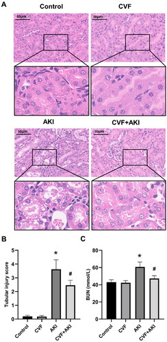
Figure 2. Evaluation of renal C3 and markers for all three complement pathways in the model of wasp venom-induced AKI.
Analysis of changes in C3, C1q, FB, and MBL in the kidneys of mice in the control, AKI, and CVF + AKI groups using immunofluorescence.
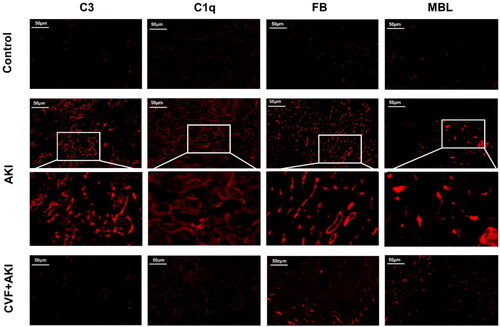
Figure 3. Complement factors C3 and C5 in kidneys were activated through all three pathways in the model of wasp venom-induced AKI.
Western blot was used to quantify C3, C5, C1q, FB, and MBL protein levels in the kidneys. *p < 0.05 versus control group, #p < 0.05 versus AKI group.
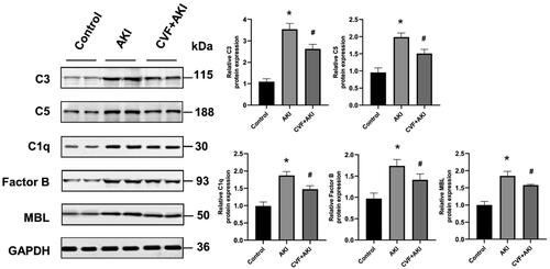
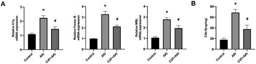
Figure 4. Assessment of renal C1q, FB, MBL and C5b-9 in the model of wasp venom-induced AKI.
(A) The mRNA expression levels of C1q, FB, and MBL in the kidneys of the three groups. (B) C5b-9 was measured using ELISA in three groups. *p < 0.05 versus control group, #p < 0.05 versus AKI group.