
Figures & data
Table 1. Clinical characteristics of Normal group and PE group.
Figure 1. Endonuclease G (ENDOG) was highly expressed in preeclampsia (PE) tissues and cells. A: ENDOG was differentially expressed according to Venn diagram analysis in PE- and hypoxia-related datasets. In GSE147776, the expression of ENDOG in the PE group (n = 7) was significantly higher than that in the normal group (n = 8). B–D: ENDOG was highly expressed in placental tissues collected from patients with PE, as demonstrated by the results of western blotting, qRT-PCR, and immunohistochemistry. E: Transfection efficiency was determined using qRT-PCR. **p < 0.01 compared to the normal and vector groups; ##p < 0.01 compared to the si-control group.
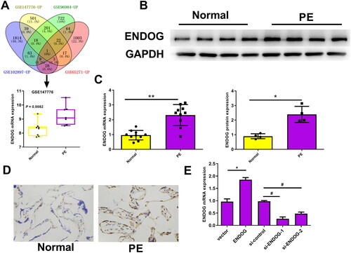
Figure 2. Endonuclease G (ENDOG) regulated HTR-8/SVneo cell activity and proliferation. A: Cell viability was determined using the CCK-8 assay. B: Cyclin A, PCNA, and CDK1 protein levels were determined using western blotting. C: Cell proliferation was determined using the EdU assay. **p < 0.01 indicates a significant difference compared to the vector group; ##p < 0.01 indicates a significant difference compared to the si-control group.
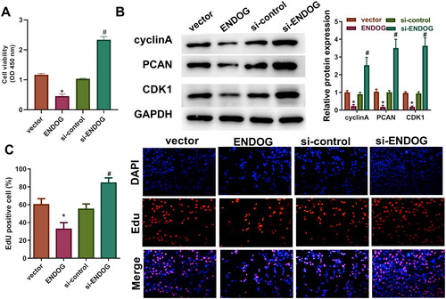
Figure 3. Endonuclease G (ENDOG) regulated HTR-8/SVneo cell invasion and migration. A: Cell invasion and migration were detected using a transwell assay. B: Cell migration was determined using a wound healing assay. C: E-cadherin, N-cadherin, and vimentin levels were determined using western blotting. **p < 0.01 indicates a significant difference compared to the vector group; ##p < 0.01 indicates a significant difference compared to the si-control group.
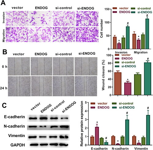
Figure 4. Endonuclease G (ENDOG) regulated angiogenesis in HTR8/SVneo cells. A: Tube formation was determined using the tube formation assay. B: ELISA was performed to determine the concentration of angiotensin (Ang)-1. C: VEGFA, PUMA, Bcl-2, Bax, TRIF, and ENDOG levels were determined using western blotting. **p < 0.01 compared to the vector group; #p < 0.05 compared to the si-control group.
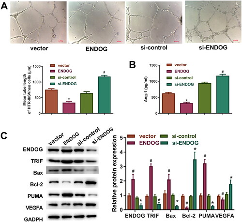
Figure 5. Endonuclease G (ENDOG) regulated the Wnt signaling pathway in HTR-8/SVneo cells. A and B: KEGG pathways enriched with differentially expressed genes (DEGs). C: Western blotting was used to determine the levels of Wnt7B, Wnt5B, β-catenin, and C-myc. **p < 0.01 compared to the vector group; ##p < 0.01 compared to the si-control group.
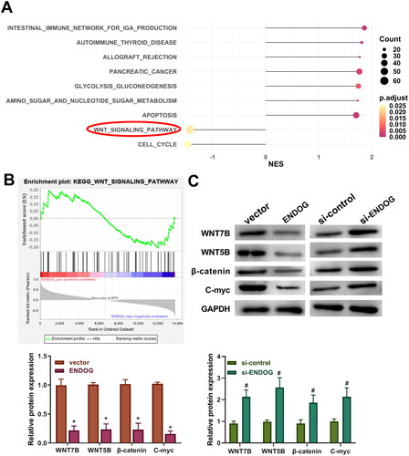
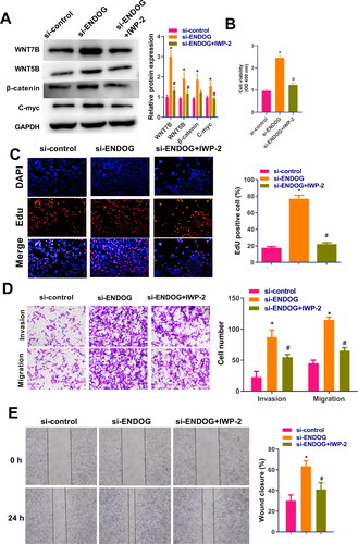
Figure 6. Endonuclease G (ENDOG) regulated HTR-8/SVneo cell activity, proliferation, invasion, and migration via activation of the Wnt signaling pathway. A: Western blotting was used to determine the levels of Wnt7B, Wnt5B, β-catenin, and C-myc. B: Cell viability was measured using the CCK-8 assay. C: Cell proliferation was measured using the EdU assay. D: Cell viability, invasion, and migration were measured using the transwell assay. E: Cell migration was measured using the wound healing assay. **p < 0.01 compared to the si-control group; ##p < 0.01 compared to the si-ENDOG group.
Supplemental Material
Download MS Word (87.3 KB)Data availability statement
The datasets used and analyzed during the current study are available from the corresponding author on reasonable request.