
Figures & data
Figure 1. Circ_0129657 was upregulated in OGD-induced HBMECs. (A) The expression of circ_0129657 was assessed by RT-qPCR. (B and C) The stability of circ_0129657 was tested by Rnase R and actinomycin D treatments in OGD-induced HBMECs. (D) The formation and structure of circ_0129657 were shown. ***p < 0.001, n = 3.
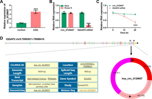
Figure 2. Silencing circ_0129657 promoted cell proliferation and inhibited apoptosis and inflammatory factor expression in OGD-induced HBMECs. (A-M) HBMECs were introduced with si-circ_0129657 or si-NC before OGD treatment. (A) Circ_0129657 knockdown efficiency was detected by RT-qPCR. (B) Effect of silencing circ_0129657 on cell viability was assessed by CCK-8 assay. (C) Changes in cell proliferation were detected after silencing circ_0129657 by EdU assay. (D) The caspase-3 activity was investigated by ELISA kits. (E) The effect of silencing circ_0129657 on apoptosis was measured by flow cytometry assay in OGD-induced HBMECs. (F–I) The protein expression levels of PCNA, Bax and Bcl2 were assessed by western blot assay after silencing circ_0129657 expression. (J–M) The protein expression levels of IL-1β, IL-6 and TNF-α were investigated by western blot assay in OGD-induced HBMECs. ***p < 0.001, n = 3.
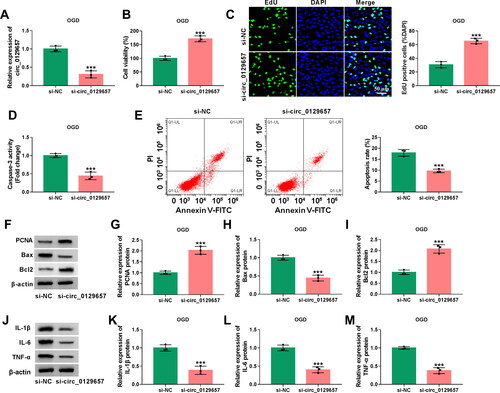
Figure 3. Circ_0129657 directly bound to miR-194-5p. (A) The putative miR-194-5p binding of circ_0129657 was shown. (B) The expression of miR-194-5p was measured by RT-qPCR in OGD-induced HBMECs. (C) The luciferase activity of circ_0129657WT and circ_0129657MUT was detected by dual-luciferase reporter assay. (D and E) RIP and RNA pull-down assays were used to assess the interaction between circ_0129657 and miR-194-5p. ***p < 0.001, n = 3.
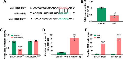
Figure 4. MiR-194-5p inhibitor partially rescued the effects of circ_0129657 silencing on cell biological properties in OGD-induced HBMECs. (A) The knockdown efficiency of miR-194-5p was examined by RT-qPCR in HBMECs after transfection by anti-miR-194-5p or anti-miR-NC. (B-M) HBMECs were introduced with si-circ_0129657 + anti-miR-194-5p, si-circ_0129657 + anti-miR-NC, si-circ_0129657 or si-NC before OGD treatment. (B) Cell viability was assessed by CCK-8 assay. (C) EdU assay was used to measure cell proliferative capacity. (D and E) Caspase-3 activity and apoptosis of cells were assessed by ELISA kits and flow cytometry assay, respectively. (F-I) The western blot assay examined the protein expression of PCNA, Bax and Bcl2 in OGD-induced HBMECs after the indicated transfection. (J-M) Western blot assay was used to assess the expression of IL-1β, IL-6 and TNF-α. **p < 0.01, ***p < 0.001, n = 3.
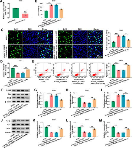
Figure 5. MiR-194-5p targeted GMFB. (A) The binding site of miR-194-5p and GMFB was shown. (B and C) The mRNA expression and protein expression of GMFB were assessed by RT-qPCR and western blot assay. (D) Dual-luciferase reporter assay was used to measure the luciferase activity of GMFB-3′UTRWT or GMFB-3′UTRMUT after co-transfection with miR-194-5p mimic or miR-NC mimic. (E) The protein expression of GMFB was tested after transfection with miR-194-5p inhibitor by western blot assay. (F) Western blot assay was used to check the protein expression of GMFB after transfection with si-NC, si-circ_0129657, si-circ_0129657 + anti-miR-NC or si-circ_0129657 + anti-miR-194-5p in OGD-induced HBMECs. **p < 0.01, ***p < 0.001, n = 3.
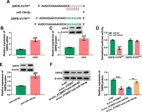
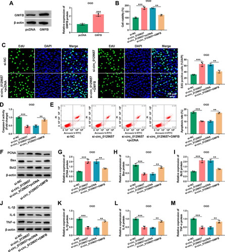
Figure 6. The influence of circ_0129657 and GMFB on OGD-induced HBMECs progression. (A) The protein expression of GMFB was detected using western blot assay in OGD-induced HBMECs after transfection by GMFB expression plasmid or pcDNA control. (B-M) HBMECs were transfected with si-NC, si-circ_0129657, si-circ_0129657 + pcDNA or si-circ_0129657 + GMFB expression plasmid. (B) CCK-8 assay was used to analyze the viability of cells treated as indicated. (C) EdU assay was used to investigate cell proliferation. (D) ELISA kits were used to detect caspase-3 activity in treated HBMECs. (E) Flow cytometry assay was used to assess the apoptosis rate of treated HBMECs. (F–I) The expression levels of PCNA, Bax and Bcl2 were tested by western blot assay. (J–M) Western blot assay was used to measure the protein expression of IL-1β, IL-6 and TNF-α. **p < 0.01, ***p < 0.001, n = 3.
Supplemental Material
Download MS Word (1.5 MB)Data availability statement
The datasets used and analyzed during the current study are available from the corresponding author on reasonable request.