Figures & data
Figure 1. LAMP3 is up-regulated in irradiation-resistant HNSCC cells. (A) LAMP3 expression pattern in HNSC tissue samples and normal samples was obtained from GEPIA database. Red asterisk represents p value less than 0.01. (B) LAMP3 expression in five HNSCC cell lines (SCC9, SCC15, FaDu, CAL-27, HN30) in comparison with human immortalised normal mucosal cell line DOK was measured by RT-qPCR. (C) Cell Counting Kit-8 assay was applied to verify the viability of irradiation-resistant HNSCC cell lines (FaDu/IR and CAL-27/IR) and irradiation-sensitive HNSCC cell lines (FaDu/S and CAL-27/S). (D) The survival rate of cells were treated with different units of irradiation (0, 2, 4, 6, 8 Gy) was evaluated through colony formation assay. (E) LAMP3 expression in irradiation-resistant HNSCC cells and sensitive cells was measured by RT-qPCR. **P < 0.01.
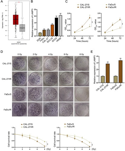
Figure 2. LAMP3 enhances radioresistance of HNSCC cells. (A) LAMP3 expression was overexpressed in CAL-27/S cells while was silenced in CAL-27/IR cells, as demonstrated by RT-qPCR. (B) The viability of CAL-27/S cells upon the ectopic expression of LAMP3 as well as that of CAL-27/IR cells with LAMP3 silence was analysed by cell counting kit-8 assay. (C) The proliferation abilities of LAMP3-overexpressed CAL-27/S cells and LAMP3-silenced CAL-27/IR cells were evaluated by colony formation assay. (D-E) Transwell assays were performed to verify the impact of LAMP3 overexpression (or knockdown) on the migration and invasion of CAL-27/S cells (or CAL-27/IR cells). (F) The protein levels of EMT-related markers (E-cadherin, N-cadherin, Vimentin) in CAL-27/S cells transfected with LAMP3 overexpression vector or CAL-27/IR cells transfected with LAMP3-specific shRNAs, as measured by western blot. **P < 0.01.
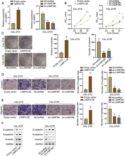
Figure 3. LAMP3 induces the autophagy of HNSCC cells. (A) The expression of autophagy-related proteins (Beclin1, p62, LC3I/II) in CAL-27/S transfected with LAMP3 overexpression vector and CAL-27/IR cells LAMP3-specific shRNAs was analysed by western blot. (B) Immumofluorescence was utilised to measure the fluorescence intensity of anti-LC3 labelled by red fluorescence (named RFP-LC3) or green fluorescence (named GFP-LC3). (C) The apoptosis of CAL-27/S transfected with LAMP3 overexpression vector and CAL-27/IR cells LAMP3-specific shRNAs was assessed by flow cytometry. (D) TUNEL assay was taken to evaluate the apoptosis of CAL-27/S transfected with LAMP3 overexpression vector and CAL-27/IR cells LAMP3-specific shRNAs. **P < 0.01.
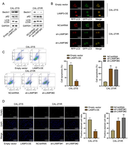
Figure 4. Irradiation-resistant HNSCC cells transfers exosomal LAMP3 into irradiation-sensitive HNSCC cells. Irradiation-sensitive HNSCC cells co-cultured with irradiation-resistant HNSCC cells were named CAL-27/IR-CM and FaDu/IR-CM groups. The normally cultured irradiation-sensitive HNSCC cells were taken as control groups. (A) Cell counting kit-8 assay was taken to verify the viability of sensitive cells before or after co-culturing. (B) The survival rate of sensitive cells treated with different units of radiation (0, 2, 4, 6, 8 Gy) was analysed by colony formation assays before or after co-culturing. (C) LAMP3 expression was measured in CAL-27/IR-CM and FaDu/IR-CM and control sensitive cells by RT-qPCR. (D) The existence of exosomes in CAL-27/IR and FaDu/IR cells was detected using electron microscope. (E) PKH67 green fluorescence staining was used to label exosomes secreted by CAL-27/IR and FaDu/IR cells. (F) Exosome surface markers (CD9, CD63, TSG101) expression in CAL-27/IR-exo and FaDu/IR-exo and the cell lysate of CAL-27/IR and FaDu/IR cells, as detected by western blot. (G) Cell counting kit-8 assay was taken to measure the viability of irradiation-sensitive HNSCC cells under exosome treatment. (H) Colony formation assay was taken to detect the proliferation of irradiation-sensitive HNSCC cells under exosome treatment. (I) The expression level of LAMP3 in sensitive HNSCC cells was measured by RT-qPCR after treated with CAL-27/IR-exo or FaDu/IR-exo. (J) The effect of CAL-27/IR-exo and FaDu/IR-exo on the promoter activity of LAMP3 in CAL-27/S and FaDu/S cells was measured by luciferase reporter assay. **P < 0.01.
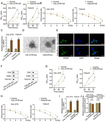
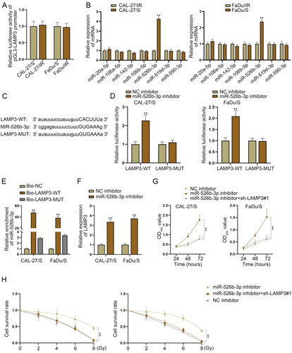
Figure 5. MiR-526b-3p suppresses LAMP3 expression to enhance radiosensitivity of HNSCC-sensitive cells. (A) Luciferase reporter assay was applied to measure the promoter activity of LAMP3 in resistant and sensitive cells. (B) Expression of miRNAs potentially interacted with LAMP3 in irradiation-resistant FaDu/IR and CAL-27/IR cells and irradiation-sensitive FaDu/S and CAL-27/S cells was measured via RT-qPCR. (C) The binding sites between miR-526b-3p and LAMP3 3’UTR as well as the mutant sequence of LAMP3. (D) The wild type sequence of LAMP3 3’UTR and the mutant one were separately inserted into pmirGLO reporter vector, and then co-transfected into sensitive HNSCC cells along with miR-526b-3p inhibitor and NC inhibitor. The firefly luciferase activity of each vector was measured by a dual luciferase reporter assay system. (E) The enrichment of miR-526b-3p was detected in the pulldown products of Bio-LAMP3-WT, Bio-LAMP3-MUT and Bio-NC by RNA pulldown assay. (F) LAMP3 expression was measured in CAL-27/S and CAL-27/IR cells after miR-526b-3p silencing. (G) Cell counting kit-8 assay was taken to measure the viability of sensitive cells transfected with NC inhibitor, miR-526b-3p inhibitor or miR-526b-3p inhibitor + sh-LAMP3#1. (H) Colony formation assay was carried out in sensitive cells transfected with NC inhibitor, miR-526b-3p inhibitor or miR-526b-3p inhibitor + sh-LAMP3#1 to measure their survival rate under different dose of irradiation. **P < 0.01.