
Figures & data
Figure 1. circFLNA was elevated in patients with ARDS and mice following CLP operation. (A) circFLNA expression analyses accessed by RT-qPCR in BALF of patients with ARDS and non-ARDS controls (n = 10). (B) ROC curve of circFLNA in sepsis-induced ARDS (n = 10). (C) Comparison of convergent and divergent primer amplifications in cDNA and gDNA of circFLNA (n = 3). (D). circFLNA schematic diagram of shear. (E) mRNA expression of circFLNA and its linear form before and after digestion by rnase R (n = 3). (F) RT-qPCR analysis of circFLNA and its linear form mRNA expression by actinomycin D treatment (n = 3). (G) pathological changes of lung tissues from ARDS mice and sham group were observed in HE staining (n = 6). RT-qPCR analyses of circFLNA levels in lung tissues (H) and BALF (I) of ARDS mice model (n = 3). ***p < 0.001. circ: cyclic RNA; ARDS: acute respiratory distress syndrome; CLP: cecal ligation and puncture; BALF: bronchoalveolar lavage fluid; ROC: receiver operator characteristic; BALF, bronchoalveolar lavage fluid.
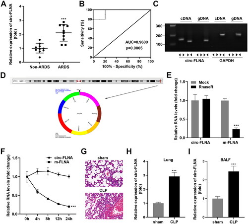
Figure 2. Depletion circFLNA ameliorated inflammatory response of sepsis-induced ARDS mice. (A) the RT-qPCR analyze of circFLNA expression in HEK293T cells transfected with LV-sh-circFLNA plasmids (n = 3). (B) Two-parameter frequency profile of flow cytometry (n = 6). (C) the percentages of CD4+CD25+Foxp3+ Tregs in different groups (n = 6). (D–F) TNF-α, IL-1β, and IL-6 concentrations of lung tissues obtained from mice were accessed by ELISA (n = 6).**p < 0.01, ***p < 0.001, vs. sham group. ##p < 0.01, vs. CLP group. LV: lentivirus; circ: cyclic RNA; TNF-α: tumor necrosis factor-α; IL-1β: interleukin 1 beta; IL-6: interleukin 6; CLP: cecal ligation and puncture; NC: negative control; sh: short hairpin RNA.
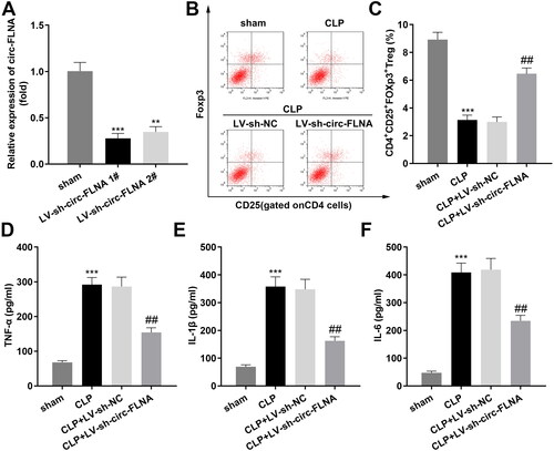
Figure 3. miR-214-5p Serves as a target of circFLNA. (A) Binding sites between miR-214-5p and circFLNA. (B) Luciferase activity, and (C) RNA pull-down assays were performed to verify the binding relationship (n = 3). (D) RT-qPCR analyses of miR-214-5p expression in BALF of sepsis-induced ARDS patients (n = 10). RT-qPCR analyses of miR-214-5p expression in lung tissues (E) and BALF (F) (n = 6). (G) RT-qPCR analyses of miR-214-5p expression in HEK293T cells transfected with inhibited circFLNA (n = 3). ***p < 0.001, vs. mimic NC, biotin-NC, non-ARDS, sham, and LV-sh-NC groups. circ: cyclic RNA; WT, wild type; MUT, wild type; NC: negative control; ARDS: acute respiratory distress syndrome; CLP: cecal ligation and puncture; BALF, bronchoalveolar lavage fluid.
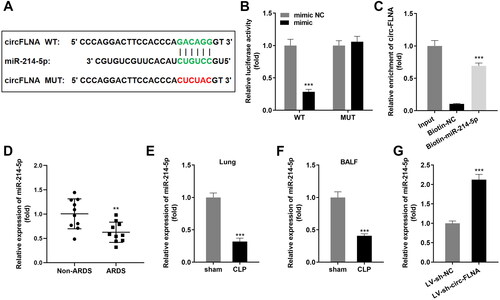
Figure 4. Inhibition of miR-214-5p abrogated the functions of inhibited circFLNA on inflammatory response of sepsis-induced ARDS mice. (A) the RT-qPCR analyze of miR-214-5p expression in HEK293T cells before and after transfection (n = 3). (B) Two-parameter frequency profile of flow cytometry (n = 6). (C) The percentages of CD4+CD25+Foxp3+ Tregs in different groups (n = 6). (D–F). TNF-α, IL-1β, and IL-6 concentrations of lung tissues obtained from mice were accessed by ELISA (n = 6). ***p < 0.001, vs. control group. ##p < 0.01, vs. CLP group. &&p < 0.01, vs. CLP + LV-sh-circFLNA + AntagomiR-NC group. LV: lentivirus; circ: cyclic RNA; TNF-α: tumor necrosis factor-α; IL-1β: interleukin 1 beta; IL-6: interleukin 6; CLP: cecal ligation and puncture; NC: negative control; sh: short hairpin RNA.
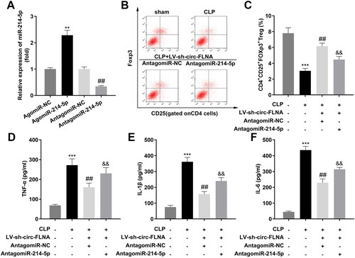
Figure 5. PD-1 is a target gene of miR-214-5p. (A) The binding sites of miR-214-5p and PD-1 were predicted. (B) Luciferase activity and (C) RNA pull-down assays were performed to verify the binding relationship (n = 3). (D) RT-qPCR analyses of PD-1 expression in BALF of sepsis-induced ARDS patients (n = 10). RT-qPCR analyses of PD-1 expression in lung tissues (E) and BALF (F) (n = 6). (G) RT-qPCR analyses of PD-1 expression in HEK293T cells transfected with upregulated miR-214-5p (n = 3). ***p < 0.001, vs. mimic NC, Biotin-NC, Non-ARDS, sham, and AgomiR-NC groups. circ: cyclic RNA; WT, wild type; MUT, wild type; NC: negative control; ARDS: acute respiratory distress syndrome; CLP: cecal ligation and puncture; BALF, bronchoalveolar lavage fluid.
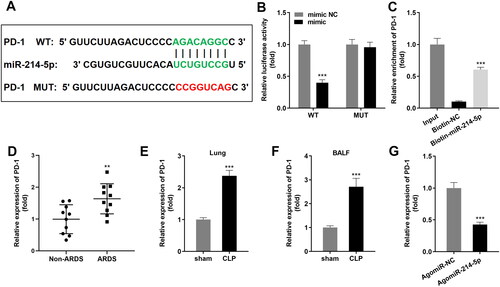
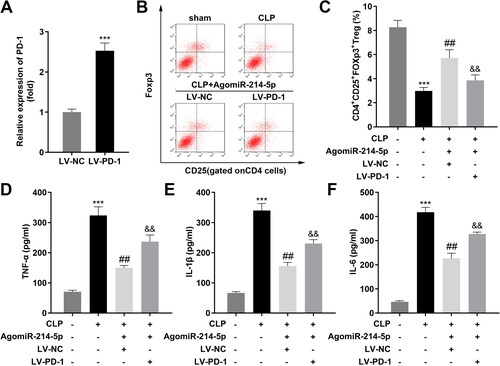
Figure 6. Over-expression of PD-1 inhibited the effects of upregulated miR-214-5p. (A) The RT-qPCR analyze of PD-1 expression in HEK293T cells before and after transfection (n = 3). (B) Two-parameter frequency profile of flow cytometry (n = 6). (C) The percentages of CD4 + CD25 + Foxp3+ Tregs in different groups (n = 6). (D–F) TNF-α, IL-1β, and IL-6 concentrations of lung tissues obtained from mice were accessed by ELISA (n = 6). ***p < 0.001, vs. control group. ##p < 0.01, vs. CLP group. &&p < 0.01, vs. CLP + AgomiR-214-5p + LV-NC group. LV: lentivirus; TNF-α: tumor necrosis factor-α; IL-1β: interleukin 1 beta; IL-6: interleukin 6; CLP: cecal ligation and puncture; NC: negative control; sh: short hairpin RNA.
Data availability statement
The datasets used and analyzed during the current study are available from the corresponding author on reasonable request.