
Figures & data
Figure 1. DUSP12 expression in OGD/R-exposed neurons and overexpression of DUSP12 by adenovirus. The expression of DUSP12 in HT22 neurons post-OGD/R was examined by (A) qRT-PCR and (B, C) Western blotting. (D, E) HT22 neurons were infected with Ad-DUSP12 or Ad-Ctrl for 48 h and DUSP12 expression was detected by Western blotting. n = 3. **p < 0.01. Statistical differences were determined using Student’s t-test.
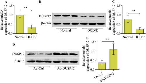
Figure 2. The effect of DUSP12 overexpression on neuronal OGD/R injury. Ad-Ctrl- or Ad-DUSP12-infected HT22 neurons were used to establish the OGD/R model and (A, B) DUSP12 expression was examined by Western blotting. (C) The viability of Ad-Ctrl- or Ad-DUSP12-infected neurons with or without OGD/R was measured by the Calcein AM cell viability assay. The apoptosis of Ad-Ctrl- or Ad-DUSP12-infected neurons with or without OGD/R was assessed by (D, E) TUNEL (scale bar = 100 μm) and (F, G) Annexin V-FITC/PI staining assays. (H, I) Levels of Bcl2, Bax and BDNF in Ad-Ctrl- or Ad-DUSP12-infected neurons with or without OGD/R were detected by Western blotting. n = 3. *p < 0.05, **p < 0.01, and ***p < 0.001. Statistical differences were determined using one-way ANOVA followed by Tukey’s post-hoc test.
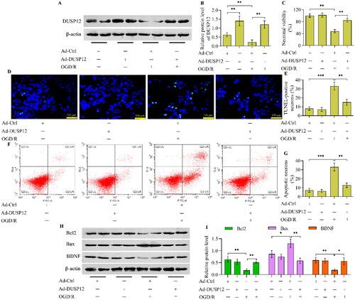
Figure 3. The effect of DUSP12 overexpression OGD/R-evoked inflammatory response. (A-C) Concentrations of TNF-α, IL-6 and IL-1β in Ad-Ctrl- or Ad-DUSP12-infected neurons with or without OGD/R were quantified by ELISA. (D, E) Levels of phosphorylated NF-κB p65 in Ad-Ctrl- or Ad-DUSP12-infected neurons with or without OGD/R were examined by Western blotting. n = 3. **p < 0.01, and ***p < 0.001. Statistical differences were determined using one-way ANOVA followed by Tukey’s post-hoc test.
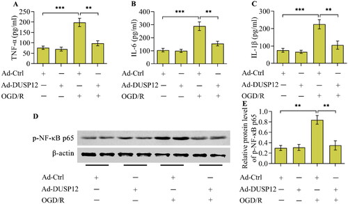
Figure 4. The effect of DUSP12 deficiency on neuronal OGD/R injury. (A, B) DUSP12 expression in Ad-sh-Scrambled- or Ad-sh-DUSP12-infected HT22 neurons with or without OGD/R was examined by Western blotting. (C) The viability of Ad-sh-Scrambled- or Ad-sh-DUSP12-infected neurons with or without OGD/R was assessed by the Calcein AM cell viability assay. The apoptosis of Ad-sh-Scrambled- or Ad-sh-DUSP12-infected neurons with or without OGD/R was evaluated by the (D, E) TUNEL (scale bar = 100 μm) and (F, G) Annexin V-FITC/PI staining assays. (H, I) Levels of Bcl2, Bax and BDNF in Ad-sh-Scrambled- or Ad-sh-DUSP12-infected neurons with or without OGD/R were examined by Western blotting. n = 3. *p < 0.05, **p < 0.01, and ***p < 0.001. Statistical differences were determined using one-way ANOVA followed by Tukey’s post-hoc test.
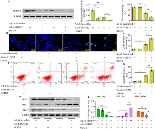
Figure 5. The effect of DUSP12 deficiency on the OGD/R-evoked inflammatory response. (A-C) Concentrations of TNF-α, IL-6 and IL-1β in Ad-sh-Scrambled- or Ad-sh-DUSP12-infected neurons with or without OGD/R were quantified by ELISA. (D, E) Levels of phosphorylated NF-κB p65 in Ad-sh-Scrambled- or Ad-sh-DUSP12-infected neurons with or without OGD/R were detected by Western blotting. n = 3. **p < 0.01, and ***p < 0.001. Statistical differences were determined using one-way ANOVA followed by Tukey’s post-hoc test.
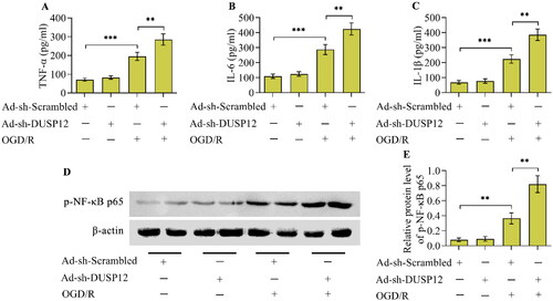
Figure 6. The effect of DUSP12 overexpression on ASK1-JNK/p38 MAPK under OGD/R conditions. (A-D) The levels of phosphorylated ASK1, JNK and p38 in Ad-Ctrl- or Ad-DUSP12-infected neurons with or without OGD/R were determined by Western blotting and their protein quantification was shown. n = 3. **p < 0.01. Statistical differences were determined using one-way ANOVA followed by Tukey’s post-hoc test.
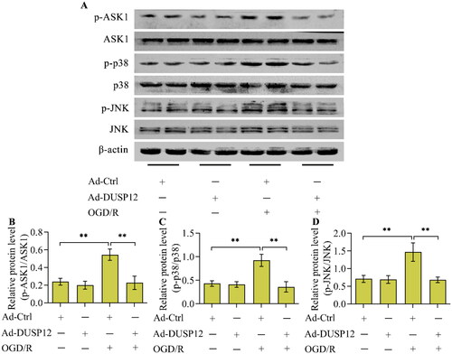
Figure 7. The effect of DUSP12 deficiency on ASK1-JNK/p38 MAPK under OGD/R conditions. (A-D) The levels of phosphorylated ASK1, JNK and p38 in Ad-sh-Scrambled- or Ad-sh-DUSP12-infected neurons with or without OGD/R were determined by Western blotting and their protein quantification was shown. n = 3. **p < 0.01. Statistical differences were determined using one-way ANOVA followed by Tukey’s post-hoc test.
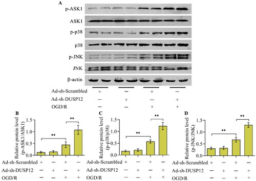
Figure 8. The effect of ASK1 inhibition on DUSP12-mediated JNK/p38 MAPK. Ad-sh-DUSP12-infected neurons were treated with the ASK1 inhibitor GS-4997 and subjected to OGD/R. (A, D) Levels of DUSP12 and phosphorylated ASK1, JNK and p38 were examined by Western blotting and their protein quantification was shown. n = 3. **p < 0.01 and #p > 0.05. Statistical differences were determined using one-way ANOVA followed by Tukey’s post-hoc test.
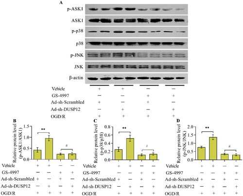
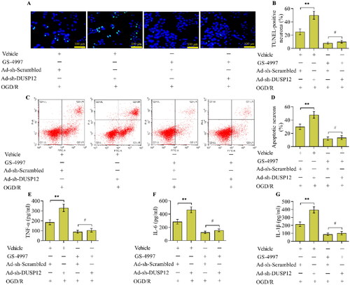
Figure 9. The effect of ASK1 inhibition on DUSP12-mediated OGD/R injury. Neuronal apoptosis in different groups was monitored by the (A, B) TUNEL (scale bar = 100 μm) and (C, D) Annexin V-FITC/PI staining assays. (E, F) Concentrations of TNF-α, IL-6 and IL-1β in different groups were quantified by ELISA. n = 3. **p < 0.01 and #p > 0.05. Statistical differences were determined using one-way ANOVA followed by Tukey’s post-hoc test.
Data availability statement
The datasets used in this study are available from the corresponding author upon reasonable request.