Figures & data
Table 1. The primer sequences used for RT-PCR and gene cloning.
Fig. 1. Expression pattern of miR-185 during 3T3-L1 cell differentiation.
Notes: (A) Morphological changes in 3T3-L1 cells between day 0 and day 8 of differentiation using Oil Red O staining; (B) after Oil Red O staining, the lipid content of cultured cells was assessed by the optical density (OD) value measured at 510 nm on a spectrophotometer; (C) the expression level of miR-185 was examined during 3T3-L1 cell differentiation under normal culture conditions. The results are presented as mean ± SEM; n = 3 (*p < 0.05, **p < 0.01).
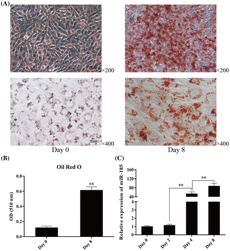
Fig. 2. MiR-185 overexpression inhibits 3T3-L1 cell differentiation.
Notes: 3T3-L1 cells were transfected with miR-185 agomirs or negative control (NC) at a density of 80–90% and differentiation was induced with DM after contact inhibition. (A) MiR-185 expression was measured by stem-loop qPCR on day 2, 4, and 8 after DM induction; photomicrograph (B) and quantitative Oil Red O (C) analysis showed that miR-185 overexpression attenuated lipid accumulation; (D–G) mRNA expression of peroxisome proliferator-activated receptor gamma (PPARγ), fatty acid binding protein 4 (FABP4), fatty acid synthase (FAS) and lipoprotein lipase (LPL) were determined by RT-PCR on day 2, 4, and 8 after DM induction. Results are expressed as fold abundance relative to NC cells on day 0; (H and I) representative western blots of whole-cell lysates at day 8 showing that PPARγ and FAS protein expression decreased in agomir-transfected cells. β-actin was used as a loading control. Results are presented as mean ± SEM; n = 3 (*p < 0.05, **p < 0.01).
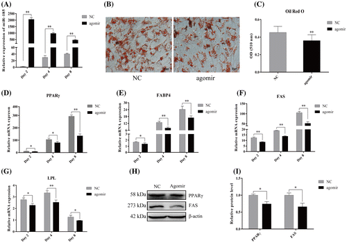
Fig. 3. MiR-185 antagomism promotes 3T3-L1 cells differentiation.
Notes: 3T3-L1 cells were transfected with miR-185 antagomirs or the corresponding negative control (NC) at a density of 80–90% and differentiation was induced with DM after contact inhibition. (A) The expression of miR-185 was measured by stem-loop qPCR on day 2, 4, and 8 after DM induction; photomicrograph (B) and quantification of Oil Red O (C) showed that miR-185 suppression increased lipid accumulation; (D–G) mRNA expression of PPARγ, FABP4, FAS and LPL were determined by RT-PCR on day 2, 4, and 8 after DMI induction. Results are expressed as fold abundance relative to NC (day 0) cells; (H and I) Representative western blots of whole cell lysates from day 8 showing that the protein expression of PPARγ and FAS increased in antagomir-transfected cells. β-actin was used as a loading control. Results are presented as mean ± SEM; n = 3 (*p < 0.05, **p < 0.01).
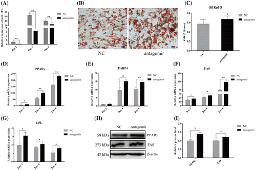
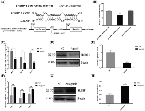
Fig. 4. MiR-185 directly targets the 3′ UTR of SREBP-1 to regulate 3T3-L1 cell differentiation.
Notes: (A) RNAhybrid-predicted binding site of miR-185 in the 3′ UTR of SREBP-1, and a schematic of luciferase reporter vector construct; (B) pmirGLO or pmirGLO-SREBP-1 3′ UTR was co-transfected with miR-185 agomirs or NC into Hela cells, and normalized Firefly luciferase activity was determined; SREBP-1 mRNA expression after transfection with miR-185 agomirs (C) or antagomirs (F) was examined by RT-PCR on day 2, 4 and 8 after DM induction. SREBP-1 protein levels on day 8 were analyzed by western blotting (D and G) and quantitative determination (E and H). β-actin was used as a loading control. The results are presented as mean ± SEM; n = 3 (*p < 0.05, **p < 0.01).