248
Views
0
CrossRef citations to date
0
Altmetric
Original Article

Adalimumab Dose Reduction and Withdrawal in Stable Non-Infectious Pediatric Uveitis: An Open-Label, Prospective, Pilot Study

, MD, , MD, PhD, , MD, PhD, , MD, , MD, PhD, , MD & , MD, PhDORCID Icon show all
Received 05 Oct 2023, Accepted 09 Apr 2024, Published online: 23 Apr 2024

Figures & data

Figure 1. An adalimumab dose reduction and withdrawal protocol by extending the dosing interval. (Adalimumab 20 mg for the patient less than 30 Kg and 40 mg for those over 30 Kg).
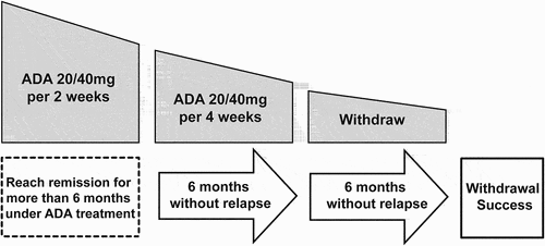
Figure 1. An adalimumab dose reduction and withdrawal protocol by extending the dosing interval. (Adalimumab 20 mg for the patient less than 30 Kg and 40 mg for those over 30 Kg).

Table 1. Demographic characteristics of children with stable non-infectious uveitis.

Figure 2. Kaplan-Meier survival curves showing relapse-free survival of all stable NIU patients after ADA dose reduction and withdrawal during the follow-up time. n = patients.
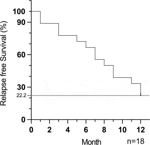
Figure 2. Kaplan-Meier survival curves showing relapse-free survival of all stable NIU patients after ADA dose reduction and withdrawal during the follow-up time. n = patients.

Table 2. Clinical and treatment features of non-infectious uveitis children before and after adalimumab-dose reduction and withdrawal.

Figure 3. Change of BCVA (A) and CMT (B) after ADA dose reduction and withdrawal during the follow-up time. BCVA: best corrected visual acuity, CMT = central macular thickness. n = eyes.
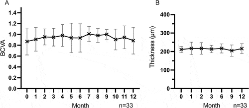
Figure 3. Change of BCVA (A) and CMT (B) after ADA dose reduction and withdrawal during the follow-up time. BCVA: best corrected visual acuity, CMT = central macular thickness. n = eyes.

Figure 4. Change of anterior chamber cell (ACC) grade (A) and vitritis grade (B) after ADA dose reduction and withdrawal during the follow-up time. n = eyes.

Figure 4. Change of anterior chamber cell (ACC) grade (A) and vitritis grade (B) after ADA dose reduction and withdrawal during the follow-up time. n = eyes.

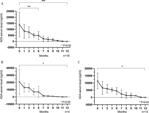
Figure 5. (A) All patients’ ADA serum trough level gradually decreased during follow-up, and decreased to 0 at the last visit. (B,C) The ADA serum trough level decreased gradually to 0 over time in both the non-relapse group (B) and relapse group (C) n = patients.

Figure 5. (A) All patients’ ADA serum trough level gradually decreased during follow-up, and decreased to 0 at the last visit. (B,C) The ADA serum trough level decreased gradually to 0 over time in both the non-relapse group (B) and relapse group (C) n = patients.

Data availability statement

The data that support the findings of this study are available from the corresponding author upon reasonable request.