Figures & data
Figure 1. Specific IgY antibody ELISA values in the egg yolk from chickens immunised with Sigma gliadin (200 µg/ml protein) in PBS, emulsified with Freund's incomplete adjuvant. Booster immunisations were given at 2 and 4 weeks after the initial immunisation. Values are the mean of quadruple samples, with vertical bars indicating the standard deviation.
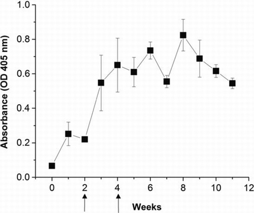
Figure 2. The properties of water soluble egg yolk (absorbance at 280 nm) and IgY activity (ELISA) following fractionation by Sephacryl S-300 chromatography. Flow rate: 3 ml/h, Column: 1.0×110 cm. The IgY fraction containing 180 kDa was verified by SDS–PAGE.
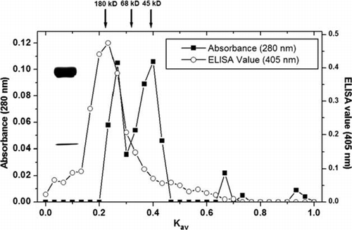
Figure 3. Standard curve of DAS-ELISA for the detection limit of 5 ng/ml of gliadin (a) and the working linear range at 10–80 ng/ml of gliadin (b). Vertical bars indicate standard deviation. Straight line indicates the linear fit ‘y=0.006x+0.201, R 2=0.99’.
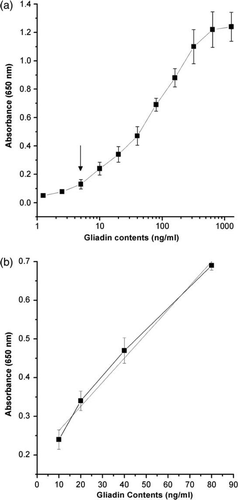
Table 1. Gliadin contents in bread samples at dilutions of 1:512 to 1:8192 in PBS from the stock sample preparation by DAS-ELISA (ng/ml; mean±SD).
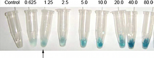
Figure 4. Immunoswab assay for detection of gliadin. Increased intensity of swab colour observed with increasing gliadin concentration (from left to right) with the control swab (left side) tested in the absence of antigen. Arrow indicates the detection limit of gliadin.