1,170
Views
0
CrossRef citations to date
0
Altmetric
Case Report

Progressive mucinous histiocytosis treated successfully with thalidomide: a rare case report

ORCID Icon, , , & ORCID Icon
Article: 2117538 | Received 20 May 2022, Accepted 22 Aug 2022, Published online: 21 Oct 2022

Figures & data

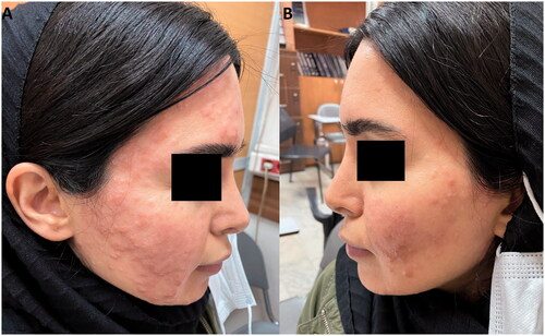
Figure 1. Erythematous papular lesions on the face.

Figure 1. Erythematous papular lesions on the face.

Figure 2. (A) H&E staining shows a dense infiltrate involving the full thickness of the dermis. There is no grenz zone between the infiltrate and surface/follicular epithelium. -The infiltrate has a vaguely storiform growth pattern (x100) and is composed of epithelioid and spindle-shaped histiocytes arranged in a collagenous stroma with mucin deposition. There is also a perivascular lymphoplasmacytic infiltration in the dermis (x400). (B) CD68; Immunohistochemistry staining for CD68 markers shows positive staining in most histiocytes. (C) CD1a; CD1a is negative in tumoral cells but positive in normal tissue Langerhans cells. (D) S100; S100 is negative in tumoral cells and highlights normal tissue dendritic cells and Langerhans cells (x100).

Figure 2. (A) H&E staining shows a dense infiltrate involving the full thickness of the dermis. There is no grenz zone between the infiltrate and surface/follicular epithelium. -The infiltrate has a vaguely storiform growth pattern (x100) and is composed of epithelioid and spindle-shaped histiocytes arranged in a collagenous stroma with mucin deposition. There is also a perivascular lymphoplasmacytic infiltration in the dermis (x400). (B) CD68; Immunohistochemistry staining for CD68 markers shows positive staining in most histiocytes. (C) CD1a; CD1a is negative in tumoral cells but positive in normal tissue Langerhans cells. (D) S100; S100 is negative in tumoral cells and highlights normal tissue dendritic cells and Langerhans cells (x100).

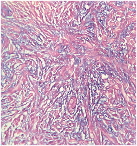
Figure 3. There is abundant mucin deposition in the dermis (alcian blue stain, x400).

Figure 3. There is abundant mucin deposition in the dermis (alcian blue stain, x400).

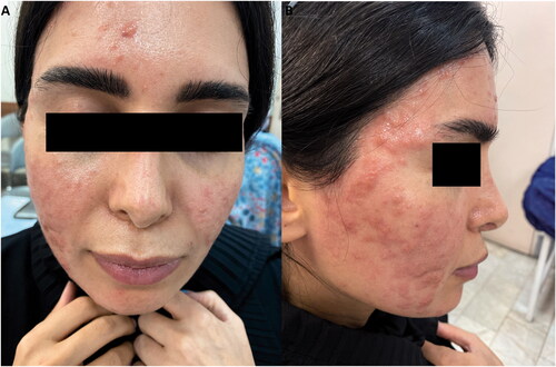
Figure 4. (A/B) Decrease in number and size of papules following one month of treatment with thalidomide.

Figure 4. (A/B) Decrease in number and size of papules following one month of treatment with thalidomide.

Table 1. Review of literature on progressive mucinous histiocytosis hereditary and sporadic cases in chronological order.