
Figures & data
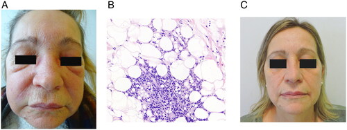
Figure 1. (A) Initial lesions the patient presented. Facial angioedema and subcutaneous erythematous nodules located on the forehead and cheeks. (B) Histological examination. Lobar panniculitis with focally granulomatous inflammation containing coalescing ‘holes’ of nonpolarizable clear material suggestive of silicone (hematoxylin–eosin, original magnification ×200). (C) Complete clearance of skin lesions after 1 months under treatment with adalimumab. No lesions have been evidenced in the follow up for the last 26 months.
Data availability statement
The data that support the findings are available from the corresponding author, JGC, upon reasonable request.