
Figures & data
Figure 1. Histopathological pattern of leukocytoclasia without vasculitis. (A) Microscopic observation of leukocytoclasia without vasculitis. Evolution of SchS before and after canakinumab treatment. (B) Skin wounds before canakinumab treatment (side view). (C) Skin wounds before canakinumab treatment (dorsal view). (D) Skin recovery after one dose of canakinumab (side view). (E) Skin recovery after one dose of canakinumab (dorsal view).
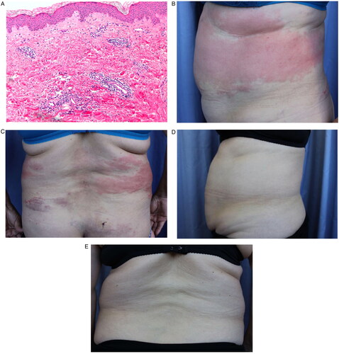
Table 1. Summary of patient characteristics with diagnosis of Schnitzler syndrome.
Data availability statement
Data sharing not applicable to this article as no datasets were generated or analyzed during the current study.