
Figures & data
Table 1. Demographic characteristics of the participants.
Figure 1. (A, B, C). Clinical photographs of three representative cases showing significant improvements in pigmentary lesions after four treatment sessions with the 785 nm picosecond Nd:YAG laser.
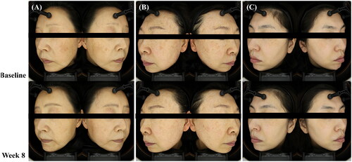
Figure 2. Evaluation of the effects of 785 nm picosecond Nd:YAG laser treatment on the (A) skin melanin index, (B) erythema index, and (C) brown index at weeks 1, 2, 3, 4, and 8 (*p < 0.05, **p < 0.01, and ***p < 0.001 by repeated measures ANOVA and post hoc Bonferroni correction).
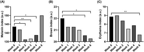
Table 2. Satisfaction of the participants.
Data availability statement
The data that support the findings of this study are available from the corresponding author, PKY, upon reasonable request.