
Figures & data
Figure 1. DFSP. Proliferation of uniform spindle cells in the dermis, separated from epidermis by a border zone. (Courtesy: Dr. Daja Šekoranja).
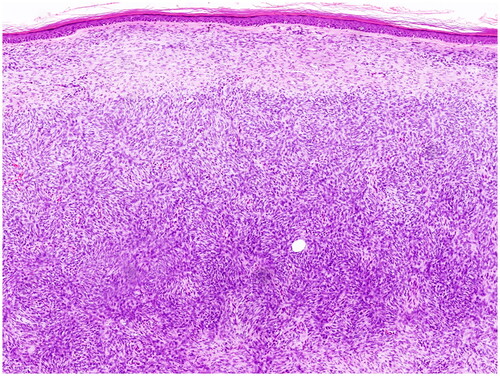
Figure 2. PRISMA flow chart.
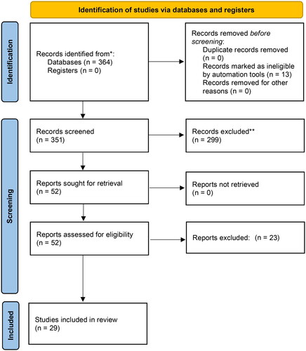
Figure 3. Rate of recurrence of DFSP in head and neck area after Mohs micrographic surgery. Case series.
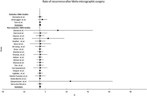
Table 1. Non-comparative studies of the treatment of DFSP of the head and neck with Mohs micrographic surgery (MMS).
Figure 4. Comparison of rate of recurrence of DFSP in head and neck area after Mohs micrographic surgery. MMS: Mohs micrographic surgery; WLE: wide local excision.
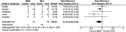
Table 2. Comparative studies between wide local excision (WLE) and Mohs micrographic surgery (MMS) for the treatment of DFSP of the head and neck.