Figures & data
Figure 1. Leptin in the epidermis and serum are higher in psoriatic patients with MS.
(A) Skin sections of normal human skin (healthy) or psoriasis without MS (without MS) and with MS lesional (with MS) skins were immunohistochemically stained with leptin antibody.
(B) The intensity of immunohistochemical staining was quantitatively rated by H-score.
(C) Serum levels of leptin in psoriatic patients without MS or those with MS were detected by ELISA.
(D) The decrease rate of PASI of psoriatic patients treated by NB-UVB for 10 days was negatively correlated with the serum leptin level (Pearson’s correlation analysis, r = −0.4022, p < 0.01, n = 62).
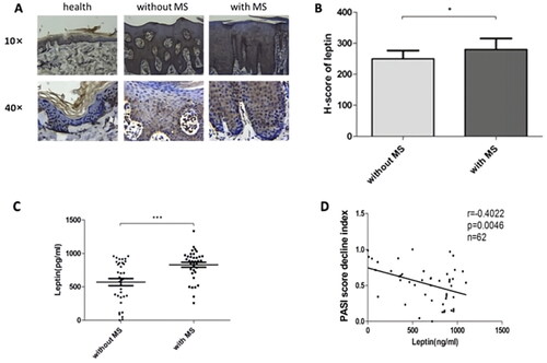
Figure 2. Induction leptin-induces insulin resistance in keratinocytes viaed by SOCS3.
(A) Starved HaCaT cells were treated with 1 μg/ml leptin for 72 hours, followed by 10 minutes of stimulation with 100 nM insulin. Immunoblotting was performed with the indicated phospho-specific or total protein antibodies.
(B) Starved HaCaT cells were treated with 1 μg/ml leptin for 48 hours and the mRNA expression of SOCS3 was determined by q-PCR.
(C) Cells were treated with 1 μg/ml leptin for 48 or 72 hours and the protein expression of SOCS3 was detected by western-blotting.
(D) HaCaT cells were transfected with 80 pmol small interfering RNA (siRNA), serum-starved, and stimulated with 1 μg/ml leptin for 72 hours, followed by 10 minutes of stimulation with 100 nM insulin. Western-blotting was conducted with the indicated phospho-specific or total protein antibodies. PKB, rotein kinase B; P-PKB, phosphorylated PKB; IRS1, insulin receptor substrates.
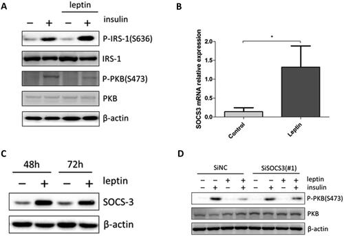
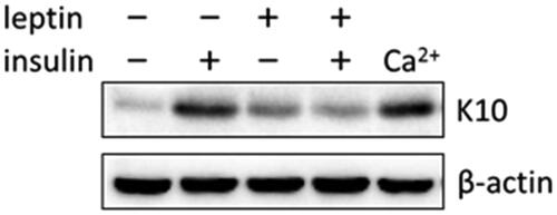
Figure 3. Leptin-induced insulin resistance interferes with the differentiation of keratinocytes. HaCaT cells were treated with 100 nM insulin, 100 ng/ml and 1 μg/ml leptin for 6 days, and the expression of keratin 10 (K10) and actin was assessed by Western blotting.