
Figures & data
Figure 1. (a,c) Clinical manifestation before treatment with tofacitinib. Multiple papules and macules on forechest and left lower limb, with most of them breaking and crusting at the top. (b,d) Clinical manifestation after treatment with upacitinib. The papules have almost faded, leaving significant hyperpigmentation.
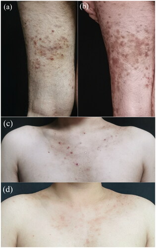
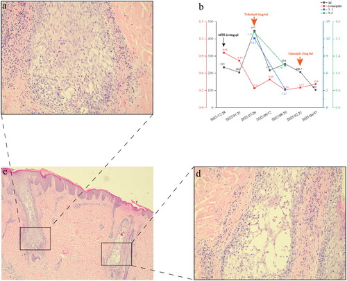
Figure 2. (a,c,d) Histopathology manifestation of the lesion obtained from the left upper arm. Hyperkeratosis, focal parakeratosis, and acanthosis. Follicular epithelium spongiosis and eosinophil infiltration were observed. Perivascular infiltrated by eosinophil and lymphocyte mildly in superficial and middle dermis (hematoxylineosin, scale bars: (a) 200×, (c) 40×, and (d) 200×). (b) Serum levels of eosinophil markers during the treatment course.
Data availability statement
All data generated or used during the case appear in the submitted manuscription.