Figures & data
Figure 1. Effect of different low dose radiation exposure regimens on the paw volume in FCA-induced arthritic rats. Rats were exposed to a total exposure level of 0.5 Gy applied either single (1 × 0.5 Gy at day 14) or split (2 × 0.25 Gy at days 14&21 or at days 21&28). Results are expressed as mean volume of edema ± SEM (n = 8/group).
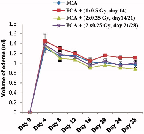
Table 1. Effect of different low dose radiation exposure regimens on the paw volume in in the Freund’s complete adjuvant-induced arthritic rats.
Table 2. Effect of different low dose radiation exposure regimens on the serum levels of pro-inflammatory cytokines and oxidative stress markers in the Freund’s complete adjuvant-induced arthritic rats.
Figure 2. Effect of low dose radiation exposure (2 × 0.25 Gy, days 14&21) and RSV (50 mg/kg) administration on the paw volume in FCA-induced arthritic rats. RSV and diclofenac were given orally for two weeks started from day 14. Results are expressed as mean volume of edema ± SEM (n = 8/group). @p < .05 compared to FCA group.
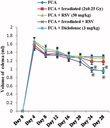
Table 3. Effect of low dose radiation exposure and resveratrol administration on the paw volume in the Freund’s complete adjuvant-induced arthritic rats.
Figure 3. Effect of low dose radiation exposure (2 × 0.25 Gy, days 14&21) and RSV (50 mg/kg) administration on the serum levels of TNF-α (a) and IL-1β (b) in FCA-induced arthritic rats. RSV and diclofenac were given orally for two weeks started from day 14. Results are expressed as mean ± SEM (n = 8/group). #p < .05 compared to Normal group. @p < .05 compared to FCA group. *p < .05 compared to irradiated group.
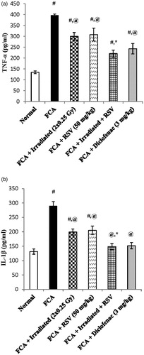
Figure 4. Effect of low dose radiation exposure (2 × 0.25 Gy, days 14&21) and RSV (50 mg/kg) administration on the serum levels of TBARs (a) and NOx (b) in FCA-induced arthritic rats. RSV and diclofenac were given orally for two weeks started from day 14. Results are expressed as mean ± SEM (n = 8/group). #p < .05 compared to Normal group. @p < .05 compared to FCA group. *p < .05 compared to irradiated group.

Figure 5. Representative sections of ankle joint stained with hematoxylin and eosin (H&E magnification x200) of (a) Normal group, (b) FCA group, (c) FCA + Irradiated (2 × 0.25 Gy, days 14&21) group, (d) FCA + RSV (50 mg/kg) group, (e) FCA + Irradiated + RSV group and (f) FCA + diclofenac (3 mg/kg) group. (g) Tissue sections were scored individually for infiltration of inflammatory cells (large arrow), synovial proliferation, pannus formation (small arrow) and cartilage damage (arrow head) on a scale 0–3. (h) Total histological scoring of joints was evaluated and the sum of all mean scores was calculated. (i) Number of inflammatory cells/microscopic field. Results are expressed as mean ± SEM (n = 8/group). #p < .05 compared to Normal group. @p < .05 compared to FCA group. *p < .05 compared to irradiated group.
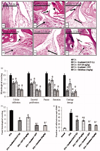
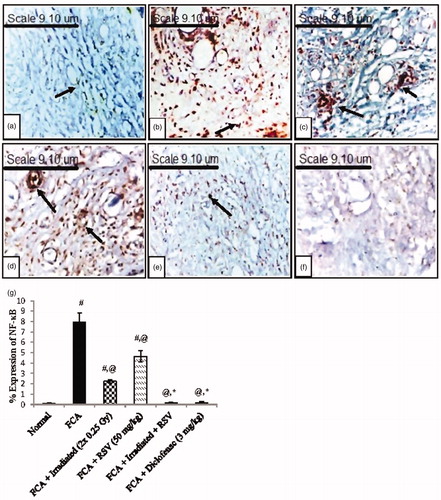
Figure 6. Immuno-histochemical analysis (magnification x400) of the NF-κB p65 expression in synovial tissues of (a) Normal group, (b) FCA group, (c) FCA + irradiated (2 × 0.25 Gy, days 14&21) group, (d) FCA + RSV (50 mg/kg) group, (e) FCA + irradiated + RSV group and (f) FCA + diclofenac (3 mg/kg) group. (g) Quantitative analysis of NF-κB p65 expression in synovial tissues of normal, arthritic and treated groups. Results are expressed as mean percentage of expression area ± SEM (n = 8/group). #p < .05 compared to Normal group. @p < .05 compared to FCA group. *p < .05 compared to irradiated group.