Figures & data
Table 1. List of rbcL gene and ITS sequences used in this study.
Figs. 1–7 Light microscopy of an aplanosporic strain of Eudorina unicocca originating from Lake Tsukui, Kanagawa, Japan (TKI-C-2). . Surface view of 32-celled vegetative colony. Note a single, basal pyrenoid (P) in the chloroplast of each cell. . Optical section of 32-celled vegetative colony. Arrowheads indicate colonial envelope surrounding whole colony. . Surface view of vegetative cell, showing contractile vacuoles (arrowheads) distributed in the protoplast surface. . Optical section of vegetative cell, showing a single large pyrenoid (P) in the chloroplast, and contractile vacuoles (arrowheads) distributed in the protoplast surface. . Daughter colony formation in asexual reproduction. Note each cell is enclosed by a transparent vesicle or cellular envelope (arrows) within the parental gelatinous matrix (arrowheads). . Vegetative colony stained with methylene blue. Individual cellular sheaths of the colonial gelatinous matrix (asterisks) are apparent. . Mature aplanospores 27 days after transferring into nitrogen-deficient medium.
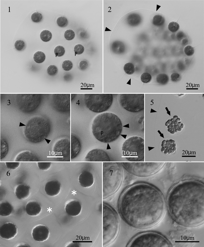
Fig. 8. Line drawing of 32-celled vegetative colony of an aplanosporic strain of Eudorina unicocca originating from Lake Tsukui, Kanagawa Prefecture, Japan.
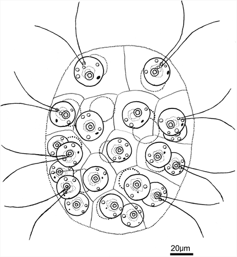
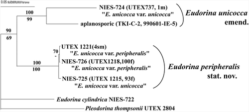
Fig. 9. Neighbor-joining (NJ) tree based on rbcL genes from 17 strains of Eudorina species, 17 strains of Volvox, five strains of Pleodorina, one strain of Platydorina, eight strains of Yamagishiella, and two strains of Pandorina (). Branch lengths are propotional to Kimura (Citation1980) distances, which are indicated by the scale bar besides the tree. Numbers above or below the branches represent 50% or more bootstrap values based on 1,000 replications of the NJ or maximum parsimony analyses, respectively.
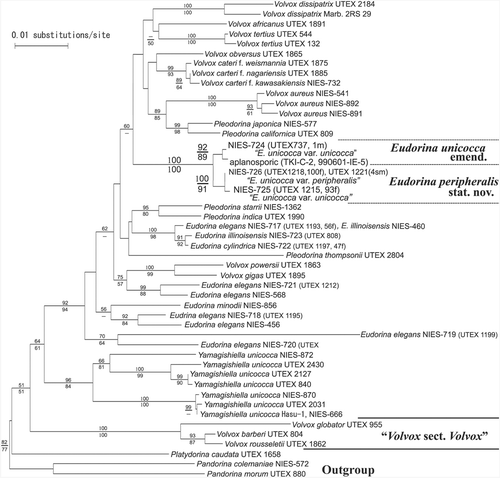
Fig. 10. Neighbor-joining (NJ) tree based on ITS sequences from 6 strains of Eudorina species and ones strain of Pleodorina (). Branch lengths are proportional to Kimura (Citation1980) distances, which are indicated by the scale bar besides the tree. Numbers above or below the branches represent 50% or more bootstrap values based on 1,000 replications of the NJ or maximum parsimony analyses, respectively.
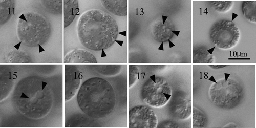
Figs 11–18. Vegetative cells of Eudorina and Yamagoshiella showing two types of distribution of contractile vacuoles in the protoplasts. . Eudorina. Note each cell has several contractile vacuoles (arrowheads) distributed in the protoplast surface. , give surface views of protoplasts and , provide optical sections. , . E. unicocca NIES-724 (UTEX 737, 1m). . E. peripheralis NIES-725 (UTEX 1215, 93f). . E. illinoisensis NIES-723. . Yamagishiella unicocca. Note only two anterior contractile vacuoles (arrowheads) in the protoplast surface. , , are surface views of anterior regions of protoplasts and an optical section of the protoplast. No contractile vacuoles are visible in the periphery. , . Hasu-2. . NIES-762. . NIES-870.
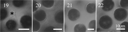
Figs. 19–22. Vegetative colonies of Eudorina unicocca and E. peripheralis stained with methylene blue. . E. unicocca NIES-724 (UTEX 737,1m), showing individual cellular sheaths (asterisk). . E. peripheralis NIES-725 (UTEX 1215, 93f). . E. peripheralis NIES-726 (UTEX 1218, 100f). . E. peripheralis UTEX 1221 (4sm). Scale bars: 10µm.