Figures & data
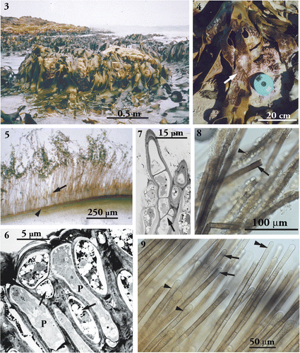
Fig. 1. Phylogenetic tree of the Phaeophyceae based on the maximum likelihood analysis of combined partial rbcL and 26S data (GTR+I+G model, –ln likelihood=14013.8779). Numbers above lines indicate bootstrap values (left: ML; right: MP), numbers below Bayesian posterior probabilities. Dashes indicate that branches received a bootstrap support of 50% or less, or less than 70% Bayesian probability. The scale bar indicates substitutions per site.

Fig. 2. Phylogenetic tree (ML) for basal Phaeophyceae inferred from partial rbcL sequences (GTR+I+G model, –ln likelihood=−8376.330601). Numbers above lines indicate bootstrap values (left: ML; right: MP), numbers below Bayesian posterior probabilities. Dashes indicate that branches received a bootstrap support of 50% or less, or less than 70% Bayesian probability. The scale bar indicates substitutions per site.
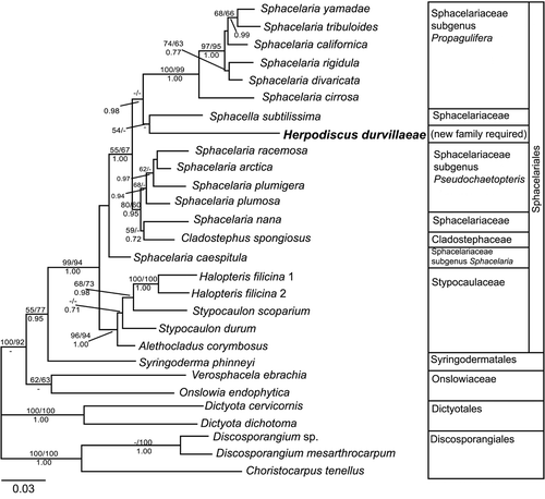
Figs. 3–9. Morphology of the parasitic of Herpodiscus durvillaeae. . Population of the host, Durvillaea antarctica, showing healthy fronds without signs of infection with H. durvillaeae. St. Kilda, 20km north of Brighton, Otago, New Zealand, March 1997. . Phylloids of the host (D. antarctica) with extensive red-brown patches of external filaments of H. durvillaeae. Brighton, August 1997. . Transverse section through external filaments of fresh H. durvillaeae Brighton, August 1997. Filaments above the host surface (arrowhead) show a row of unilocular sporangia (arrow), the tips of the filaments covered with pennate diatoms. , . Transmission electron microscopy of sections of host and parasite. . Longitudinal section through the meristoderm at the end of the host's growth season showing two apical cells of H. durvillaeae (p) among host cells at the surface. Parasite cell contents appear concentrated at their apices. Shrinkage of the host cells and the loss of most of their physodes (holes indicated by arrows) are preparation artefacts. . Longitudinal section through the base of a parasite patch. Filaments of H. durvillaeae showing acroblastic branching (arrow). , . Reaction with Eau de Javel. . Transitory blackening of cell walls of H. durvillaeae immediately after treatment (arrow). Gametangia-turned gametophytes have coloured internal walls (arrowhead). Dried material, scraped from the surface of a parasite patch. (WELT A027896). . Reaction of external terminal cells of fresh H. durvillaeae with Eau de Javel as front moves through the tips of the external filaments. Some terminal cells are completely bleached (double arrowhead), while others display blue bands of colouring just below their apical hemisphere (arrows). Terminal cells have swollen tips and contain large nuclei (arrowheads).