
Figures & data
Fig. 1. Phylogenetic tree of concatenated psbA and mt23S partial genes obtained by Bayesian inference (BI). Maximum likelihood (ML) gave the same topology. ML bootstrap values (≥ 70%) and Bayesian posterior probabilities (≥ 0.95) respectively, are indicated adjacent to the branches. Sequences for taxa in bold were generated in this study. Sequences from GenBank correspond to those included in Supplementary table S1. Scale = 0.01.
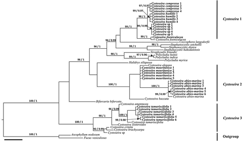
Fig. 2. Phylogenetic tree of concatenated psbA, mt23S, cox1 and nad1 partial genes obtained by Bayesian inference (BI) for the Canary Islands specimens of Cystoseira sensu lato. Maximum likelihood (ML) gave the same topology. ML bootstrap values (≥ 70%) and Bayesian posterior probabilities (≥ 0.95) respectively, are indicated adjacent to the branches. All sequences were generated in this study. Scale = 0.01.
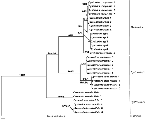
Table 1. Comparison of morphological attributes exhibited by species of Cystoseira sensu lato studied. Groups: Cystoseira 1 (= Cystoseira sensu stricto), Cystoseira 2 (here proposed as Treptacantha) and Cystoseira 3 (here proposed as Carpodesmia)
Table 2. Diameter (µm) of cells in cross section of primary branches, and diameter (mm) of reproductive structures of species of Cystoseira sensu lato
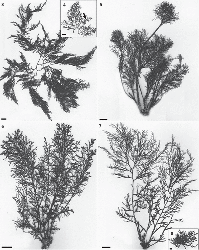
Figs 3–8. Habit of species of Cystoseira 1 (= Cystoseira sensu stricto) from the Canary Islands. Figs 3, 4. Cystoseira sp. (= C. aurantia) (TFC Phyc 15276). Fig. 3. Habit. Fig. 4. Detail of last order branches, with conspicuous aerocysts (arrows). Fig. 5. Cystoseira foeniculacea (TFC Phyc 15266). Fig. 6. Cystoseira humilis (TFC Phyc 15267). Fig. 7. Cystoseira compressa (TFC Phyc 15263). Spring-summer morphotype. Fig. 8. Cystoseira compressa (TFC Phyc 15291). Rosette morphotype. Note the caespitose habit of all species. All scale bars: 1 cm.
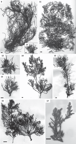
Figs 9–17. Habit of species of Cystoseira 2 (here proposed as Treptacantha). Fig. 9. Cystoseira abies-marina (TFC Phyc 15256). Well-developed specimen. Fig. 10. Cystoseira abies-marina (TFC Phyc 15259). Specimen from upper sublittoral. Fig. 11. Cystoseira usneoides (TFC Phyc 1402). Fig. 12. Cystoseira algeriensis (HGI – A 1289). Fig. 13. Cystoseira mauritanica (TFC Phyc 15271). Fig. 14. Cystoseira baccata (TFC Phyc 1766). Fig. 15. Cystoseira nodicaulis (HGI – A 3813). Fig. 16. Cystoseira elegans (HGI – A 14577). Fig. 17. Cystoseira montagnei (TFC Phyc 599). All scale bars: 1 cm.
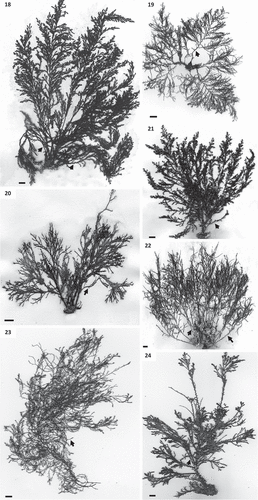
Figs 18–24. Habit of species of Cystoseira 3 (here proposed as Carpodesmia). Fig. 18. Cystoseira amentacea (HGI – A 14601). Fig. 19. Cystoseira brachycarpa (HGI – A 14552). Fig. 20. Cystoseira crinita (HGI – A 14570). Fig. 21. Cystoseira mediterranea (HGI – A 9427). Fig. 22. Cystoseira zosteroides (HGI – A 3054). Fig. 23. Cystoseira tamariscifolia (TFC Phyc 15285). Morphotype from sublittoral at La Graciosa islet. Fig. 24. Cystoseira tamariscifolia (TFC Phyc 15281). Morphotype from lower eulittoral at Gran Canaria island. Note: spiny appendages on primary branches are indicated with arrows. All scale bars: 1 cm.
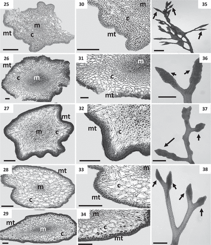
Figs 25–38. Cross sections of thalli and details of receptacles of Cystoseira 1 (= Cystoseira sensu stricto) from the Canary Islands. Figs 25–29. Primary branches. Fig. 25. Cystoseira sp. (= C. aurantia) (TFC Phyc 15276). Fig. 26. Cystoseira foeniculacea (TFC Phyc 15266). Fig. 27. Cystoseira humilis (TFC Phyc 15267). Fig. 28. Cystoseira compressa (TFC Phyc 15263). Spring-summer morphotype. Fig. 29. Cystoseira compressa (TFC Phyc 15291). Rosette morphotype. Figs 30–34. Details of the three vegetative tissues. Fig. 30. Cystoseira sp. (= C. aurantia) (TFC Phyc 15276). Fig. 31. Cystoseira foeniculacea (TFC Phyc 15266). Fig. 32. Cystoseira humilis (TFC Phyc 15267). Fig. 33. Cystoseira compressa (TFC Phyc 15263). Fig. 34. Cystoseira compressa (TFC Phyc 15291). Figs 35–38. Warty fusiform receptacles (arrows). Fig. 35. Cystoseira sp. (= C. aurantia) (TFC Phyc 15276). Fig. 36. Cystoseira foeniculacea (TFC Phyc 15266). Fig. 37. Cystoseira humilis (TFC Phyc 15267). Fig. 38. Cystoseira compressa (TFC Phyc 15263). Meristoderm (mt), cortex (c) and medulla (m). Scale bars: Figs 25–34 = 100 μm; Figs 35–38 = 1 mm.
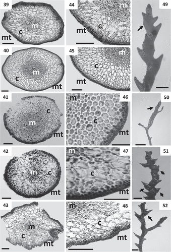
Figs 39–52. Cross sections of thalli and details of receptacles of Cystoseira 2 (here proposed as Treptacantha). Figs 39–48. Primary branches. Fig. 39. Cystoseira abies-marina (TFC Phyc 15259). Upper sublittoral morphotype. Fig. 40. Cystoseira abies-marina (TFC Phyc 15256). Sublittoral morphotype. Fig. 41. Cystoseira usneoides (TFC Phyc 1402). Fig. 42. Cystoseira algeriensis (HGI – A 1289). Fig. 43. Cystoseira mauritanica (TFC Phyc 15271). Figs 44–48. Details of the three vegetative tissues. Fig. 44. Cystoseira abies-marina (TFC Phyc 15259). Upper sublittoral morphotype. Fig. 45. Cystoseira abies-marina (TFC Phyc 15256). Sublittoral morphotype. Fig. 46. Cystoseira usneoides (TFC Phyc 1402). Fig. 47. Cystoseira algeriensis (HGI – A 1289). Fig. 48. Cystoseira mauritanica (TFC Phyc 15271). Figs 49–52. Receptacles (arrows). Fig. 49. Cystoseira abies-marina (TFC Phyc 15256). Fig. 50. Cystoseira usneoides (TFC Phyc 1402). Fig. 51. Cystoseira algeriensis (HGI – A 1289). Fig. 52. Cystoseira mauritanica (TFC Phyc 15271). Meristoderm (mt), cortex (c) and medulla (m). Scale bars: Figs 39–48 = 100 μm; Figs 49–52 = 1 mm.
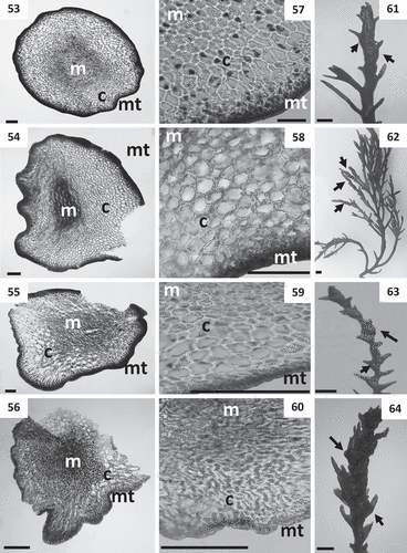
Figs 53–64. Cross sections of thalli and details of receptacles of Cystoseira 2 (continued). Figs 53–60. Primary branches. Fig. 53. Cystoseira baccata (TFC Phyc 1766). Fig. 54. Cystoseira nodicaulis (HGI – A 3813). Fig. 55. Cystoseira elegans (HGI – A 14577). Fig. 56. Cystoseira montagnei (TFC Phyc 599). Figs 57–60. Details of the three vegetative tissues. Fig. 57. Cystoseira baccata (TFC Phyc 1766). Fig. 58. Cystoseira nodicaulis (HGI – A 3813). Fig. 59. Cystoseira elegans (HGI – A 14577). Fig. 60. Cystoseira montagnei (TFC Phyc 599). Figs 61–64. Receptacles (arrows). Fig. 61. Cystoseira baccata (TFC Phyc 1766). Fig. 62. Cystoseira nodicaulis (HGI – A 3813). Fig. 63. Cystoseira elegans (HGI – A 14577). Fig. 64. Cystoseira montagnei (TFC Phyc 599). Meristoderm (mt), cortex (c) and medulla (m). Scale bars: Figs 53–60 = 100 μm; Figs 61–64 = 1 mm.
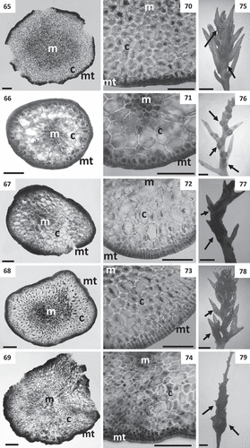
Figs 65–79. Cross sections of thalli and details of receptacles of Cystoseira 3 (here proposed as Carpodesmia). Figs 65–69. Primary branches. Fig. 65. Cystoseira amentacea (HGI – A 14601). Fig. 66. Cystoseira brachycarpa (HGI – A 14552). Fig. 67. Cystoseira crinita (HGI – A 14570). Fig. 68. Cystoseira mediterranea (HGI – A 9427). Fig. 69. Cystoseira zosteroides (HGI – A 3054). Figs 70–74. Details of the three vegetative tissues. Fig. 70. Cystoseira amentacea (HGI – A 14601). Fig. 71. Cystoseira brachycarpa (HGI – A 14552). Fig. 72. Cystoseira crinita (HGI – A 14570). Fig. 73. Cystoseira mediterranea (HGI – A 9427). Fig. 74. Cystoseira zosteroides (HGI – A 3054). Figs 75–79. Receptacles (arrows). Fig. 75. Cystoseira amentacea (HGI – A 14601). Fig. 76. Cystoseira brachycarpa (HGI – A 14552). Fig. 77. Cystoseira crinita (HGI – A 14570). Fig. 78. Cystoseira mediterranea (HGI – A 9427). Fig. 79. Cystoseira zosteroides (HGI – A 3054). Meristoderm (mt), cortex (c) and medulla (m). Scale bars: Figs 65–74 = 100 μm; Figs 75–79 = 1 mm.
Figs 80–85. Cross sections of thalli and details of receptacles of Cystoseira 3 (continued). Figs 80–83. Primary branches of Cystoseira tamariscifolia. Fig. 80. Cystoseira tamariscifolia (TFC Phyc 15285). Sublittoral morphotype. Fig. 81. Cystoseira tamariscifolia (TFC Phyc 15281). Low eulittoral morphotype. Figs 82, 83. Details of the three vegetative tissues. Fig. 82. Cystoseira tamariscifolia (TFC Phyc 15285). Sublittoral morphotype. Fig. 83. Cystoseira tamariscifolia (TFC Phyc 15281). Low eulittoral morphotype. Figs 84, 85. Receptacles (arrows). Fig. 84. Cystoseira tamariscifolia (TFC Phyc 15285). Sublittoral morphotype. Fig. 85. Cystoseira tamariscifolia (TFC Phyc 15281). Low eulittoral morphotype. Meristoderm (mt), cortex (c) and medulla (m). Scale bars: Figs 80–83 = 100 μm; Figs 84–85 = 1 mm.
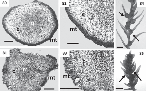
Table 3. Inferred pairwise-distances (%) for the four genetic markers studied among nearby species of Cystoseira sensu lato from the Canary Islands. Groups: Cystoseira 1 (= Cystoseira sensu stricto), Cystoseira 2 (here proposed as Treptacantha) and Cystoseira 3 (here proposed as Carpodesmia)