Figures & data
Figure 1. Fluorescence micrographs showing (discoid) human erythrocytes treated with CTB plus anti-CTB. Erythrocytes post-fixed (60 min, RT) with either PFA (5%) or GA (1%) are shown in (A) and (B), respectively. (A inset); erythrocytes incubated with CTB only. Erythrocytes post- or pre-treated with methyl-β-cyclodextrin relative to CTB plus anti-CTB treatment are shown in (C) and (C inset), respectively. Cells were fixed with PFA (5%) and GA (0.01%). Erythrocytes fixed with PFA (5%) or PFA (5%) and GA (0.01%) prior to CTB plus anti-CTB treatment are shown in (D) and (D insets), respectively.
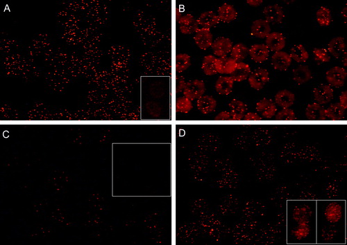
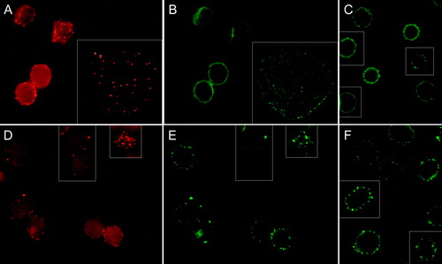
Figure 2. Fluorescence micrographs showing (discoid) human erythrocytes treated with CTB plus anti-CTB and stained for either CD47 or CD59. The CTB and CD47 signals from the same fields are indicated in (A) and (B), respectively. (C); cells stained for CD47 only. Similarly, CTB and CD59 signals from the same fields are indicated in (D) and (E), respectively. (F); cells stained for CD59 only. Cells were fixed with PFA (5%), except (D inset) and (E inset) which were fixed with PFA (5%) and GA (0.01%).
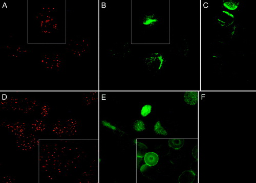
Figure 3. Fluorescence micrograph (mid-plane cross-section) with insets showing echinocytic human erythrocytes treated with CTB plus anti-CTB. Cells were incubated with A23187 (2 µM, 20 min, 37°C) plus calcium to induce echinocytosis. Cells were fixed with PFA (5%) and GA (0.01%).
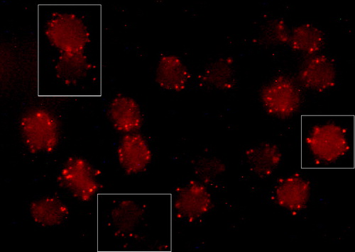
Figure 4. Fluorescence micrographs (mid-plane cross-sections) showing echinocytic human erythrocytes treated with CTB plus anti-CTB prior to staining for either CD47 or CD59. The CTB and CD47 signals from the same fields are indicated in (A) and (B), respectively. In insets spread flake-like membrane. (C); cells stained for CD47 only. Similarly, CTB and CD59 signals from the same fields are indicated in (D) and (E), respectively. (F); cells stained for CD59 only. Cells were incubated with A23187 (2 µM, 20 min, 37°C) plus calcium to induce echinocytosis. Cells were fixed with PFA (5%) and GA (0.01%).