Figures & data
Figure 1. Epithelial cells with an apical brush border. (A) Tall, columnar small intestinal enterocytes with a brush border facing the lumen of the gut. The apoptotoic cell in the middle is in the process of being extruded from the epithelium. (B) A closer view of the apical region of an enterocyte showing a dense array of microvilli with rootlets of actin filaments extending into the underlying cytoplasm. Notice that organelles such as mitochondria, lysosomes and endosomes are excluded from this uppermost terminal web region of the cytoplasm.
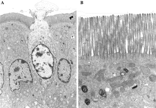
Figure 2. Bilayer membrane structure of lipid rafts. Lipid rafts prepared from intestinal microvillar membranes, using Brij 98 (A, B) or Triton X-100 (C) (Braccia et al. [Citation2003]). (A) Immunogold labeling for aminopeptidase N. Note that the labeling is confined mainly to one side of the membrane. (B, C) High magnification electron micrographs of raft membranes. The two leaflets of the bilayer are visible regardless of choice of detergent. Bars: 200 nm.
![Figure 2. Bilayer membrane structure of lipid rafts. Lipid rafts prepared from intestinal microvillar membranes, using Brij 98 (A, B) or Triton X-100 (C) (Braccia et al. [Citation2003]). (A) Immunogold labeling for aminopeptidase N. Note that the labeling is confined mainly to one side of the membrane. (B, C) High magnification electron micrographs of raft membranes. The two leaflets of the bilayer are visible regardless of choice of detergent. Bars: 200 nm.](/cms/asset/cf10f7c5-d9e3-4f03-9fd5-4ac89bb21a24/imbc_a_144543_f0002_b.jpg)
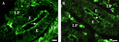
Figure 3. IgG and IgA in the enterocyte brush border. Cryosections of the crypt region of small intestinal mucosa labeled for IgG (A) or IgA (B). Both immunoglobulin classes are deposited in the brush border of enterocytes (arrows). Labeling is also seen along the basolateral surface of the enterocytes (E), as well as in plasma cells of the lamina propria (LP). Bars: 10 µm. This figure is reproduced in colour in Molecular Membrane Biology online.