326
Views
10
CrossRef citations to date
0
Altmetric
Original

Monitoring the organization and dynamics of bovine hippocampal membranes utilizing differentially localized fluorescent membrane probes

, , , , , & show all
Pages 430-441 | Received 11 Mar 2006, Published online: 09 Jul 2009

Figures & data

Figure 1.  The top panel (a) shows the chemical structures of NBD-cholesterol and NBD-PE. The lower panel (b) is a schematic representation of half of the membrane bilayer showing the localizations of the NBD groups of NBD-PE and NBD-cholesterol in phosphatidylcholine membranes. The horizontal line at the bottom indicates the center of the bilayer. Adapted and modified from reference 17.
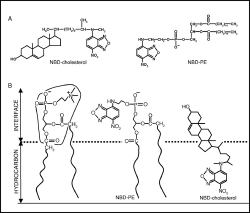
Figure 1.  The top panel (a) shows the chemical structures of NBD-cholesterol and NBD-PE. The lower panel (b) is a schematic representation of half of the membrane bilayer showing the localizations of the NBD groups of NBD-PE and NBD-cholesterol in phosphatidylcholine membranes. The horizontal line at the bottom indicates the center of the bilayer. Adapted and modified from reference 17.

Table I.  Lipid contents in native hippocampal membranes as a function of increasing cholesterol depletion.

Figure 2.  Effect of changing excitation wavelength on the wavelength of maximum emission of (a) NBD-cholesterol and (b) NBD-PE in native membranes (○), cholesterol-depleted (with 40 mM MβCD) membranes (▴) and liposomes of lipid extract from native membranes (□). The ratio of fluorophore to total phospholipid was maintained at 1:100 (mol/mol). The protein concentration is ∼0.04 mg/ml. See Materials and methods for other details.
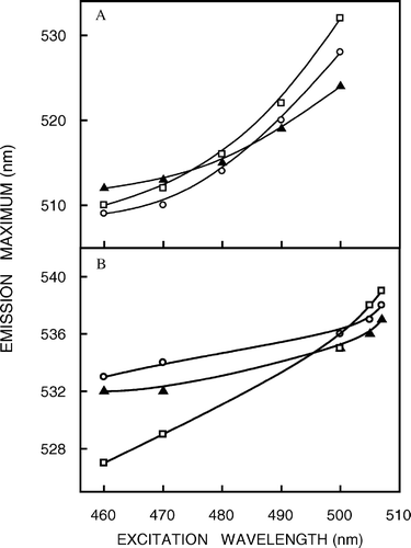
Figure 2.  Effect of changing excitation wavelength on the wavelength of maximum emission of (a) NBD-cholesterol and (b) NBD-PE in native membranes (○), cholesterol-depleted (with 40 mM MβCD) membranes (▴) and liposomes of lipid extract from native membranes (□). The ratio of fluorophore to total phospholipid was maintained at 1:100 (mol/mol). The protein concentration is ∼0.04 mg/ml. See Materials and methods for other details.

Figure 3.  (a) Effect of cholesterol depletion (by treatment with increasing concentrations of MβCD) on the magnitude of red edge excitation shift (REES) of NBD-cholesterol (○) and NBD-PE (•) in native membranes. All other conditions are as in . See Materials and methods for other details. (b) A comprehensive representation of the magnitude of red edge excitation shift (REES) obtained with NBD-cholesterol (shaded bars) and NBD-PE (white bars) in native membranes as a function of increasing cholesterol depletion (when treated with increasing concentrations of MβCD) and in liposomes of lipid extract from native membranes (data for native and cholesterol-depleted membranes taken from a). All other conditions are as in . See Materials and methods for other details.
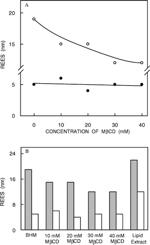
Figure 3.  (a) Effect of cholesterol depletion (by treatment with increasing concentrations of MβCD) on the magnitude of red edge excitation shift (REES) of NBD-cholesterol (○) and NBD-PE (•) in native membranes. All other conditions are as in Figure 2. See Materials and methods for other details. (b) A comprehensive representation of the magnitude of red edge excitation shift (REES) obtained with NBD-cholesterol (shaded bars) and NBD-PE (white bars) in native membranes as a function of increasing cholesterol depletion (when treated with increasing concentrations of MβCD) and in liposomes of lipid extract from native membranes (data for native and cholesterol-depleted membranes taken from Figure 3a). All other conditions are as in Figure 2. See Materials and methods for other details.

Figure 4.  Fluorescence polarization of (a) NBD-cholesterol and (b) NBD-PE in native membranes (○), cholesterol-depleted (with 40 mM MβCD) membranes (▴) and liposomes of lipid extract from native membranes (□) as a function of excitation wavelength. The emission wavelength was 512 nm for NBD-cholesterol and 531 nm for NBD-PE. All other conditions are as in . The data points shown are the means±SE of multiple measurements. See Materials and methods for other details.
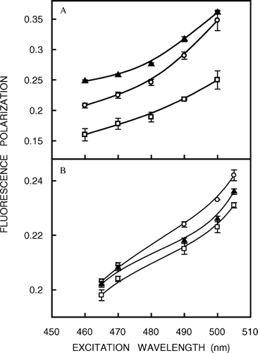
Figure 4.  Fluorescence polarization of (a) NBD-cholesterol and (b) NBD-PE in native membranes (○), cholesterol-depleted (with 40 mM MβCD) membranes (▴) and liposomes of lipid extract from native membranes (□) as a function of excitation wavelength. The emission wavelength was 512 nm for NBD-cholesterol and 531 nm for NBD-PE. All other conditions are as in Figure 2. The data points shown are the means±SE of multiple measurements. See Materials and methods for other details.

Figure 5.  (a) Effect of cholesterol depletion on the fluorescence polarization of NBD-cholesterol (○) and NBD-PE (•) in native membranes. Excitation was at 460 nm and emission was monitored at 512 nm for NBD-cholesterol while excitation was at 465 nm and emission was monitored at 531 nm for NBD-PE. The polarization data shown are the means±standard errors of multiple measurements. (b) A comprehensive representation of fluorescence polarization of NBD-cholesterol (shaded bars) and NBD-PE (white bars) in native membranes as a function of increasing cholesterol depletion and in liposomes of lipid extract from native membranes (data for native and cholesterol-depleted membranes taken from a). The data points shown are the means±SE of multiple measurements. All other conditions are as in . See Materials and methods for other details.
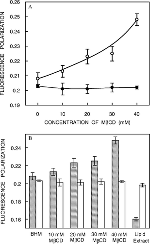
Figure 5.  (a) Effect of cholesterol depletion on the fluorescence polarization of NBD-cholesterol (○) and NBD-PE (•) in native membranes. Excitation was at 460 nm and emission was monitored at 512 nm for NBD-cholesterol while excitation was at 465 nm and emission was monitored at 531 nm for NBD-PE. The polarization data shown are the means±standard errors of multiple measurements. (b) A comprehensive representation of fluorescence polarization of NBD-cholesterol (shaded bars) and NBD-PE (white bars) in native membranes as a function of increasing cholesterol depletion and in liposomes of lipid extract from native membranes (data for native and cholesterol-depleted membranes taken from Figure 5a). The data points shown are the means±SE of multiple measurements. All other conditions are as in Figure 2. See Materials and methods for other details.

Figure 6.  Effect of cholesterol depletion on the mean fluorescence lifetime of NBD-cholesterol (○) and NBD-PE (•) in native membranes. Mean fluorescence lifetimes were calculated from using Equation 3. The data points shown are the means±SE of multiple measurements. All other conditions are as in . See Materials and methods for other details.
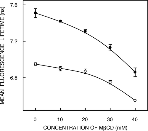
Figure 6.  Effect of cholesterol depletion on the mean fluorescence lifetime of NBD-cholesterol (○) and NBD-PE (•) in native membranes. Mean fluorescence lifetimes were calculated from Table II using Equation 3. The data points shown are the means±SE of multiple measurements. All other conditions are as in Figure 5. See Materials and methods for other details.

Table II.  Mean fluorescence lifetimes of NBD-labeled lipids in native membranes with increasing cholesterol depletion and in lipid extractsa.

Reprints and Corporate Permissions

Please note: Selecting permissions does not provide access to the full text of the article, please see our help page How do I view content?

To request a reprint or corporate permissions for this article, please click on the relevant link below:

Academic Permissions

Please note: Selecting permissions does not provide access to the full text of the article, please see our help page How do I view content?

Obtain permissions instantly via Rightslink by clicking on the button below:

If you are unable to obtain permissions via Rightslink, please complete and submit this Permissions form. For more information, please visit our Permissions help page.