409
Views
1
CrossRef citations to date
0
Altmetric
Original Articles

Evaluation of therapeutic effect of aqueous extract of stem bark of Mangifera indica Linn. on gentamicin-induced nephrotoxicity

, , &
Pages 252-256 | Received 09 Oct 2010, Accepted 13 Jan 2011, Published online: 01 Nov 2011

Figures & data

Table 1.  Effect of aqueous extract of stem bark of M. indica on SOD, lipid peroxidation, CT and RG levels.

Table 2.  Effect of aqueous extract of M. indica on serum creatinine (mg/dl) and urea (mg/dl) levels.

Table 3.  Effect of aqueous extract of M. indica on serum Na (mEq/L) and serum K (mEq/L) levels.

Figure 1.  Histopathological examination of normal group, GM group and aqueous stem bark extract of M. indica treated groups. Group 1 (H&E×100): (A) intact glomerulus; (B) intact proximal convoluted tubules.
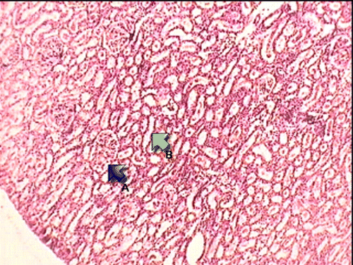
Figure 1.  Histopathological examination of normal group, GM group and aqueous stem bark extract of M. indica treated groups. Group 1 (H&E×100): (A) intact glomerulus; (B) intact proximal convoluted tubules.

Figure 2.  Histopathological examination of normal group, GM group and aqueous stem bark extract of M. indica treated groups. Group 2 (H&E×100): (A) marked glomerular changes; (B) extensive tubular necrosis; C: mononuclear cell infiltration in the interstitium.
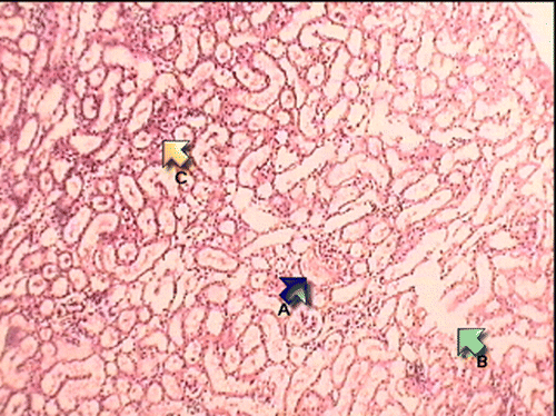
Figure 2.  Histopathological examination of normal group, GM group and aqueous stem bark extract of M. indica treated groups. Group 2 (H&E×100): (A) marked glomerular changes; (B) extensive tubular necrosis; C: mononuclear cell infiltration in the interstitium.

Figure 3.  Histopathological examination of normal group, GM group and aqueous stem bark extract of M. indica treated groups. Group 3 (H&E×400): (A) vacuolar changes in proximal convoluted tubules; (B) scattered mononuclear cell infiltration in the cortex.
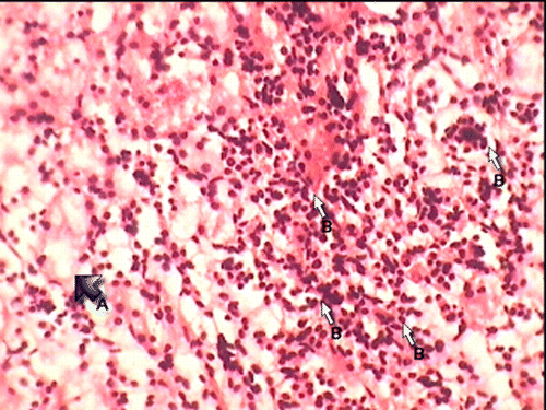
Figure 3.  Histopathological examination of normal group, GM group and aqueous stem bark extract of M. indica treated groups. Group 3 (H&E×400): (A) vacuolar changes in proximal convoluted tubules; (B) scattered mononuclear cell infiltration in the cortex.

Figure 4.  Histopathological examination of normal group, GM group and aqueous stem bark extract of M. indica treated groups. Group 4 (H&E×400): (A) mild tubular degeneration and necrosis in the subscapular area; (B) multiple hyaline cast formation in the distal convoluted tubule; (C) pronounced dilatation of distal convoluted tubules.
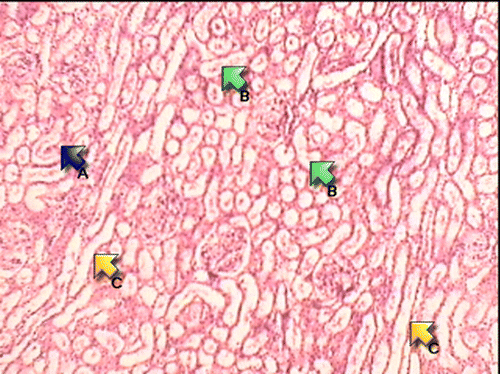
Figure 4.  Histopathological examination of normal group, GM group and aqueous stem bark extract of M. indica treated groups. Group 4 (H&E×400): (A) mild tubular degeneration and necrosis in the subscapular area; (B) multiple hyaline cast formation in the distal convoluted tubule; (C) pronounced dilatation of distal convoluted tubules.

Reprints and Corporate Permissions

Please note: Selecting permissions does not provide access to the full text of the article, please see our help page How do I view content?

To request a reprint or corporate permissions for this article, please click on the relevant link below:

Academic Permissions

Please note: Selecting permissions does not provide access to the full text of the article, please see our help page How do I view content?

Obtain permissions instantly via Rightslink by clicking on the button below:

If you are unable to obtain permissions via Rightslink, please complete and submit this Permissions form. For more information, please visit our Permissions help page.