Figures & data
Figure 1 Diagrammatic illustration of the topographical distribution of serotonergic neurons projecting to the caudate putamen and substantia nigra (blue) and serotonergic neurons projecting to brain structures implicated in the regulation of anxiety-related behavior (yellow) in rat brain (for references, see text). Although this is an oversimplification, these cell populations have a differential distribution in the dorsal raphe nucleus (DR), with serotonergic cells projecting to the caudate putamen and substantia nigra located in more rostral regions of the DR. In some cases, individual serotonergic neurons project to functionally related targets [e.g. CP/ SN (van der Kooy and Hattori Citation1980), mPFC/ Acb (Van Bockstaele et al. Citation1993)]. In some cases, serotonergic neurotransmission of the dorsal part of the mid-rostrocaudal and caudal DR can be independently regulated (Macedo et al. Citation2005), suggesting that although the innervation arises in part from the same brain region, it could also arise in part from different populations of serotonergic neurons. In the mid-rostrocaudal DR, serotonergic neurons projecting to the CP are highly concentrated within compact cell clusters in the DRD “core” and DRV “ellipse” regions. Numbers below illustrations of rostral, mid-rostrocaudal, and caudal DR indicate stereotaxic coordinates with reference to Bregma. Scale bar, 500 μm. Abbreviations, Acb, nucleus accumbens; BL, basolateral amygdala; BNST, bed nucleus of the stria terminalis; CE, central amygdaloid nucleus; CP, caudate putamen; DA, dorsal hypothalamic area; DRD, DR, dorsal part; DRI, DR, interfascicular part; DRV, DR, ventral part; DRVL, DR ventrolateral part; LC, locus coeruleus; LPB, lateral parabrachial nucleus; mPFC, medial prefrontal cortex; SN, substantia nigra.
![Figure 1 Diagrammatic illustration of the topographical distribution of serotonergic neurons projecting to the caudate putamen and substantia nigra (blue) and serotonergic neurons projecting to brain structures implicated in the regulation of anxiety-related behavior (yellow) in rat brain (for references, see text). Although this is an oversimplification, these cell populations have a differential distribution in the dorsal raphe nucleus (DR), with serotonergic cells projecting to the caudate putamen and substantia nigra located in more rostral regions of the DR. In some cases, individual serotonergic neurons project to functionally related targets [e.g. CP/ SN (van der Kooy and Hattori Citation1980), mPFC/ Acb (Van Bockstaele et al. Citation1993)]. In some cases, serotonergic neurotransmission of the dorsal part of the mid-rostrocaudal and caudal DR can be independently regulated (Macedo et al. Citation2005), suggesting that although the innervation arises in part from the same brain region, it could also arise in part from different populations of serotonergic neurons. In the mid-rostrocaudal DR, serotonergic neurons projecting to the CP are highly concentrated within compact cell clusters in the DRD “core” and DRV “ellipse” regions. Numbers below illustrations of rostral, mid-rostrocaudal, and caudal DR indicate stereotaxic coordinates with reference to Bregma. Scale bar, 500 μm. Abbreviations, Acb, nucleus accumbens; BL, basolateral amygdala; BNST, bed nucleus of the stria terminalis; CE, central amygdaloid nucleus; CP, caudate putamen; DA, dorsal hypothalamic area; DRD, DR, dorsal part; DRI, DR, interfascicular part; DRV, DR, ventral part; DRVL, DR ventrolateral part; LC, locus coeruleus; LPB, lateral parabrachial nucleus; mPFC, medial prefrontal cortex; SN, substantia nigra.](/cms/asset/89e61659-101d-4893-bd16-f492fa77f7ae/ists_a_149261_f0001_b.jpg)
Figure 2 Labeling of dorsal raphe neurons following injection of the retrograde tracer FluoroGold into the anterior part of the rat basolateral amygdala. (a) illustration of the distribution of retrogradely labeled cells within the dorsal raphe nucleus (coronal sections). Box in -8.00 mm illustration indicates the region illustrated at higher magnification in the photograph in (c). (b) photomicrograph illustrating the injection site restricted to the anterior part of the basolateral amygdaloid nucleus (-3.30 mm Bregma). (c) photomicrograph illustrating dorsal raphe neurons retrogradely labeled with FluoroGold. Retrogradely labeled dorsal raphe neurons were concentrated in the dorsal part of the mid-rostrocaudal dorsal raphe nucleus [gray shading in panel (a), levels -8.00 and -8.24 mm]. Numbers below illustrations in panel (a) indicate stereotaxic coordinates with reference to Bregma. Arrows in (c) indicate FluoroGold labeled neurons in the dorsal raphe nucleus. Abbreviations: BLA, basolateral amygdaloid nucleus, anterior part; BLP, basolateral amygdaloid nucleus, posterior part; BLV, basolateral amygdaloid nucleus, ventral part; BMP, basomedial amygdaloid nucleus, posterior part; CeC, central amygdaloid nucleus, capsular part; CeL, central amygdaloid nucleus, lateral division; LaDL, lateral amygdaloid nucleus, dorsolateral part; LaVL, lateral amygdaloid nucleus, ventrolateral part; LaVM, lateral amygdaloid nucleus, ventromedial part. Scale bar, (a) 873 μm, (b) 800 μm, (c) 50 μm.
![Figure 2 Labeling of dorsal raphe neurons following injection of the retrograde tracer FluoroGold into the anterior part of the rat basolateral amygdala. (a) illustration of the distribution of retrogradely labeled cells within the dorsal raphe nucleus (coronal sections). Box in -8.00 mm illustration indicates the region illustrated at higher magnification in the photograph in (c). (b) photomicrograph illustrating the injection site restricted to the anterior part of the basolateral amygdaloid nucleus (-3.30 mm Bregma). (c) photomicrograph illustrating dorsal raphe neurons retrogradely labeled with FluoroGold. Retrogradely labeled dorsal raphe neurons were concentrated in the dorsal part of the mid-rostrocaudal dorsal raphe nucleus [gray shading in panel (a), levels -8.00 and -8.24 mm]. Numbers below illustrations in panel (a) indicate stereotaxic coordinates with reference to Bregma. Arrows in (c) indicate FluoroGold labeled neurons in the dorsal raphe nucleus. Abbreviations: BLA, basolateral amygdaloid nucleus, anterior part; BLP, basolateral amygdaloid nucleus, posterior part; BLV, basolateral amygdaloid nucleus, ventral part; BMP, basomedial amygdaloid nucleus, posterior part; CeC, central amygdaloid nucleus, capsular part; CeL, central amygdaloid nucleus, lateral division; LaDL, lateral amygdaloid nucleus, dorsolateral part; LaVL, lateral amygdaloid nucleus, ventrolateral part; LaVM, lateral amygdaloid nucleus, ventromedial part. Scale bar, (a) 873 μm, (b) 800 μm, (c) 50 μm.](/cms/asset/0ddb3bf2-c0c6-4915-8336-5f0dfa61f64e/ists_a_149261_f0002_b.jpg)
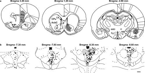
Figure 3 Diagrammatic illustration of the distribution of dorsal raphe neurons projecting to anxiety circuits. (a) illustration (coronal sections) of the location of injections of retrograde tracer into the prelimbic (PrL) and infralimbic (IL) parts of the medial prefrontal cortex (mPFC, left), nucleus accumbens (middle), and the basolateral amygdala (right). (b) illustration of the distribution of retrogradely labeled neurons in the dorsal raphe nucleus. Open circles represent single-labeled neurons following injections of retrograde tracer in the mPFC; open triangles represent single-labeled neurons following injections of retrograde tracer in the nucleus accumbens (AcbC and AcbSh regions); grey squares represent double-labeled neurons following injections of retrograde tracers into the mPFC and nucleus accumbens; black circles represent single-labeled neurons following injections of retrograde tracer in the basolateral amygdala. The dorsal part of the mid-rostrocaudal dorsal raphe nucleus (gray shading) contains concentrations of neurons with collateral projections to both the mPFC and nucleus accumbens (grey squares), and neurons projecting to the basolateral amygdala (black circles). Abbreviations: ac, anterior commissure; AOP, anterior olfactory nucleus, posterior part; BL, basolateral amygdala; AcbC, nucleus accumbens, core; AcbSh, nucleus accumbens, shell; CPu, caudate putamen; f, fornix; mPFC, medial prefrontal cortex; LV, lateral ventricle; LS, lateral septum; mt, mammillothalamic tract; opt, optic tract. Scale bar, (a) 264 μm, (b) 1 mm. Adapted from Van Bockstaele et al. Citation1993 and Abrams et al. Citation2005, with permission.