Publication Cover
Stress
The International Journal on the Biology of Stress
Volume 10, 2007 - Issue 3
1,106
Views
96
CrossRef citations to date
0
Altmetric
Original

Modulation of stress consequences by hippocampal monoaminergic, glutamatergic and nitrergic neurotransmitter systems

Review

, &
Pages 227-249 | Received 28 Sep 2006, Accepted 14 Jan 2007, Published online: 07 Jul 2009

Figures & data

Table I.  Gene expression changes in the hippocampal formation induced by stress.

Table II.  Gene expression changes in the hippocampal formation induced by antidepressant treatments.
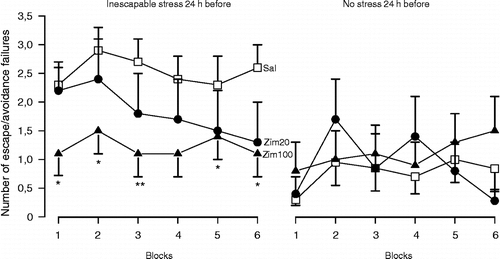
Figure 1 Post-stress facilitation of serotonin-mediated neurotransmission in the dorsal hippocampus prevents the behavioural consequences of stress. Male Wistar rats (n = 7–23) were submitted to inescapable footshocks (40 shocks, 1 mA, 10 s) or habituation (30 min) in a shuttle box. Immediately afterwards, they received bilateral intra-hippocampal injections of saline (Sal) or zimelidine (Zim, 20 or 100 nmols/0.5 μl), a selective serotonin reuptake inhibitor, and were tested 24 h later wit escapable footshocks (30 sound-signalled shocks, 0.8 mA, 10 s). Data are expressed as the mean ± SEM number of escape and/or avoidance failures in each block (summation of five individual trials). * Indicates significant difference from respective Sal-treated group (p < 0.05, ANOVA followed by Duncan test, modified from Joca et al. (Citation2003), with kind permission from Elsevier).

Figure 1 Post-stress facilitation of serotonin-mediated neurotransmission in the dorsal hippocampus prevents the behavioural consequences of stress. Male Wistar rats (n = 7–23) were submitted to inescapable footshocks (40 shocks, 1 mA, 10 s) or habituation (30 min) in a shuttle box. Immediately afterwards, they received bilateral intra-hippocampal injections of saline (Sal) or zimelidine (Zim, 20 or 100 nmols/0.5 μl), a selective serotonin reuptake inhibitor, and were tested 24 h later wit escapable footshocks (30 sound-signalled shocks, 0.8 mA, 10 s). Data are expressed as the mean ± SEM number of escape and/or avoidance failures in each block (summation of five individual trials). * Indicates significant difference from respective Sal-treated group (p < 0.05, ANOVA followed by Duncan test, modified from Joca et al. (Citation2003), with kind permission from Elsevier).

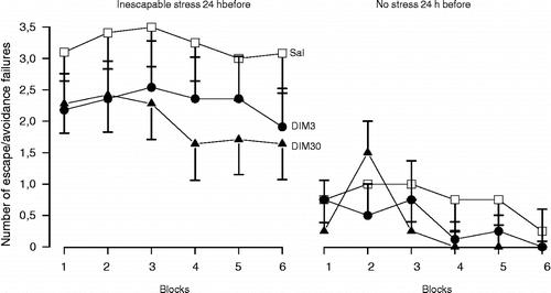
Figure 2 Post-stress facilitation of noradrenaline-mediated neurotransmission in the dorsal hippocampus does not prevent the behavioural consequences of stress. Male Wistar rats (n = 11–14) were submitted to inescapable footshocks (40 shocks, 1 mA, 10 s) or habituation (30 min) in a shuttle box. Immediately afterwards, they received bilateral intra-hippocampal injections of Sal or desipramine (DIM, 3 or 30 nmols/0.5 μl), a selective noradrenaline reuptake inhibitor and were tested 24 h later with escapable footshocks (30 sound-signalled shocks, 0.8 mA, 10 s). Data are expressed as the mean ± SEM number of escape and/or avoidance failures in each block (summation of five individual trials). There was no significant difference between treatments (modified from Joca et al. (Citation2006), with kind permission from Elsevier).

Figure 2 Post-stress facilitation of noradrenaline-mediated neurotransmission in the dorsal hippocampus does not prevent the behavioural consequences of stress. Male Wistar rats (n = 11–14) were submitted to inescapable footshocks (40 shocks, 1 mA, 10 s) or habituation (30 min) in a shuttle box. Immediately afterwards, they received bilateral intra-hippocampal injections of Sal or desipramine (DIM, 3 or 30 nmols/0.5 μl), a selective noradrenaline reuptake inhibitor and were tested 24 h later with escapable footshocks (30 sound-signalled shocks, 0.8 mA, 10 s). Data are expressed as the mean ± SEM number of escape and/or avoidance failures in each block (summation of five individual trials). There was no significant difference between treatments (modified from Joca et al. (Citation2006), with kind permission from Elsevier).

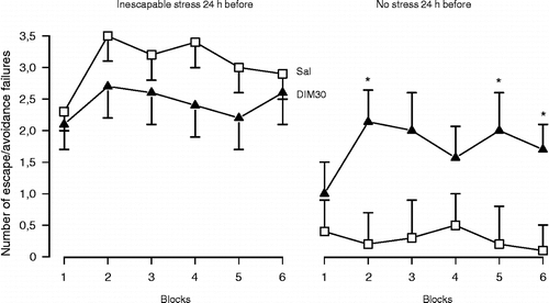
Figure 3 Pre-stress facilitation of noradrenaline-mediated neurotransmission in the dorsal hippocampus also does not prevent the behavioural consequences of stress. However, it facilitates helpless behaviour in control (non-stressed) animals. Male Wistar rats (n = 7–19) received bilateral intra-hippocampal injections of Sal or desipramine (DIM, 30 nmols/0.5 μl) and immediately afterwards were submitted to inescapable footshocks (40 shocks, 1 mA, 10 s) or habituation (30 min) in a shuttle box. All animals were tested 24 h later with escapable footshocks (30 sound-signalled shocks, 0.8 mA, 10 s). Data are expressed as the mean ± SEM number of escape and/or avoidance failures in each block (summation of five individual trials). * Indicates significant difference from respective Sal-treated group (t-test, p < 0.05, (modified from Joca et al. (Citation2006), with kind permission from Elsevier).

Figure 3 Pre-stress facilitation of noradrenaline-mediated neurotransmission in the dorsal hippocampus also does not prevent the behavioural consequences of stress. However, it facilitates helpless behaviour in control (non-stressed) animals. Male Wistar rats (n = 7–19) received bilateral intra-hippocampal injections of Sal or desipramine (DIM, 30 nmols/0.5 μl) and immediately afterwards were submitted to inescapable footshocks (40 shocks, 1 mA, 10 s) or habituation (30 min) in a shuttle box. All animals were tested 24 h later with escapable footshocks (30 sound-signalled shocks, 0.8 mA, 10 s). Data are expressed as the mean ± SEM number of escape and/or avoidance failures in each block (summation of five individual trials). * Indicates significant difference from respective Sal-treated group (t-test, p < 0.05, (modified from Joca et al. (Citation2006), with kind permission from Elsevier).

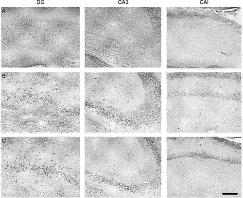
Figure 4 Subchronic treatment with a NOS inhibitor increases BDNF expression in the hippocampus. The figure shows stained cells for BDNF in the hippocampal formation after (a) vehicle, (b) imipramine (15 mg/kg) and (c) 7-nitro-indazole (60 mg/kg), a preferential nNOS inhibitor, treated rats. They received three i.p. injections (0, 5 and 24 h) and were killed 24 h after the last injection, the hippocampus was removed and processed for BDNF immunohistochemistry (primary anti-BDNF antibody: rabbit polyclonal antibody raised against a peptide mapping at the N-terminus of BDNF of human origin, Santa Cruz Biotechnology, Santa Cruz, CA, USA; 1:800). Note BDNF signal (dark stained cells) only in (b) and (c). DG, dentate gyrus; CA, ammons horn. Bar = 150 μm.

Figure 4 Subchronic treatment with a NOS inhibitor increases BDNF expression in the hippocampus. The figure shows stained cells for BDNF in the hippocampal formation after (a) vehicle, (b) imipramine (15 mg/kg) and (c) 7-nitro-indazole (60 mg/kg), a preferential nNOS inhibitor, treated rats. They received three i.p. injections (0, 5 and 24 h) and were killed 24 h after the last injection, the hippocampus was removed and processed for BDNF immunohistochemistry (primary anti-BDNF antibody: rabbit polyclonal antibody raised against a peptide mapping at the N-terminus of BDNF of human origin, Santa Cruz Biotechnology, Santa Cruz, CA, USA; 1:800). Note BDNF signal (dark stained cells) only in (b) and (c). DG, dentate gyrus; CA, ammons horn. Bar = 150 μm.

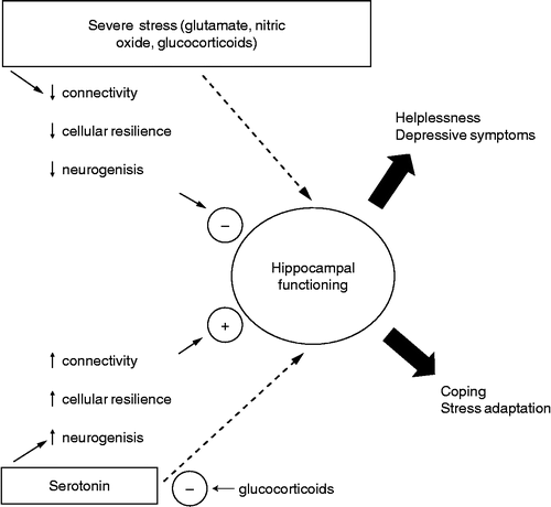
Figure 5 Severe stress, activating the NMDA/NO pathway, can impair normal hippocampal functioning, predisposing to helplessness and depressive symptoms. Conversely, serotonin can counteract the stress-induced damage to the hippocampus, restoring normal functioning and allowing coping and stress adaptation. These effects are probably mediated by 5-HT1A receptors. GCs have a permissive role in NMDA damaging effects and at high levels, can impair 5-HT1A-mediated neurotransmission. Clinical studies using tryptophan depletion and results obtained with direct intra-hippocampal drug injections suggest that serotonin, glutamate and NO can also interfere with hippocampal function independently of cellular remodelling and neurogenesis.

Figure 5 Severe stress, activating the NMDA/NO pathway, can impair normal hippocampal functioning, predisposing to helplessness and depressive symptoms. Conversely, serotonin can counteract the stress-induced damage to the hippocampus, restoring normal functioning and allowing coping and stress adaptation. These effects are probably mediated by 5-HT1A receptors. GCs have a permissive role in NMDA damaging effects and at high levels, can impair 5-HT1A-mediated neurotransmission. Clinical studies using tryptophan depletion and results obtained with direct intra-hippocampal drug injections suggest that serotonin, glutamate and NO can also interfere with hippocampal function independently of cellular remodelling and neurogenesis.

Reprints and Corporate Permissions

Please note: Selecting permissions does not provide access to the full text of the article, please see our help page How do I view content?

To request a reprint or corporate permissions for this article, please click on the relevant link below:

Academic Permissions

Please note: Selecting permissions does not provide access to the full text of the article, please see our help page How do I view content?

Obtain permissions instantly via Rightslink by clicking on the button below:

If you are unable to obtain permissions via Rightslink, please complete and submit this Permissions form. For more information, please visit our Permissions help page.