Figures & data
Table I. Subject matching and brain serotonin turnover.
Figure 1 Patterns of cerebral venous sinus drainage demonstrated by cerebral sinus scans with technetium-99, with images (posterior view) at 10–16 s after peripheral venous injection of tracer. Top, drainage of the superior sagittal sinus (carrying the bulk of cortical venous return) is primarily into the left internal jugular vein (“left dominant” pattern of drainage). Subcortical venous flow is primarily to the right internal jugular vein. Bulbar venous return (not demonstrated in the scan) is to the spinal veins. Middle, drainage of the superior sagittal sinus is largely into the right internal jugular vein “right dominant”). Subcortical flow is to the left jugular vein. Bottom, non-lateralizing venous sinus flow, with symmetrical drainage of the superior sagittal sinus into the right and left internals jugular veins, due to admixture of blood at the confluence of the sinuses.
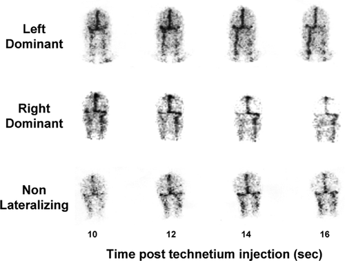
Figure 2 5-HIAA overflow from the brain into the internal jugular veins characterised on the venous sinus scan in terms of whether this was from a predominantly cortical or subcortical field of venous drainage. In those cases where the cerebral venous drainage was non-lateralizing due to vascular interconnection at the confluence of the transverse sinuses, regionalized analysis of brain serotonin turnover was not possible. Brain serotonin turnover was increased approximately 4-fold in both cortical and subcortical brain regions in panic disorder. SI conversion; ng/min = 5.23 pmol/min. **P < 0.01.
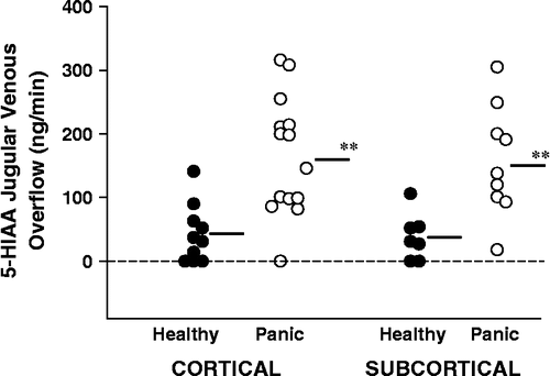
Table II. Influence of serotonin transporter gene promoter region insertion/deletion. Polymorphism on brain serotonin turnover in panic disorder.
Figure 3 The relation of severity of panic disorder, scored with the patient self-rating panic disorder severity scale, to whole brain serotonin turnover (r = 0.86, P < 0.001). SI conversion; ng/min = 5.23 pmol/min.
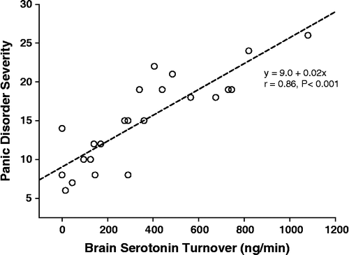
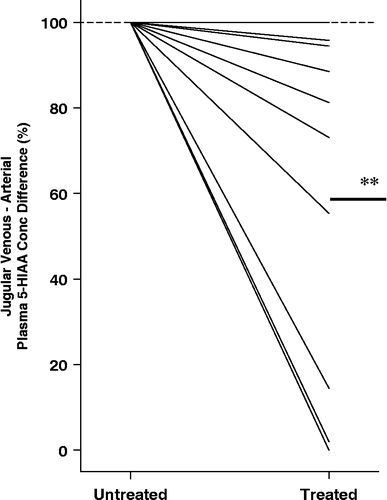
Figure 4 The effect of SSRI dosing in panic disorder, with citalopram alone or in combination with CBT, on brain serotonin turnover. The baseline veno–arterial 5-HIAA plasma concentration difference is listed as 100%, with treated serotonin turnover shown as a percentage of the initial value. A fall was seen in 9 of 10 patients (**P < 0.01).