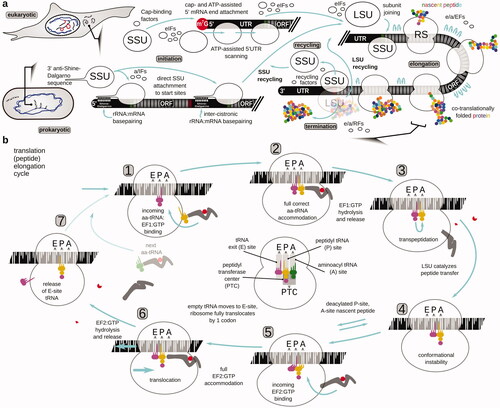
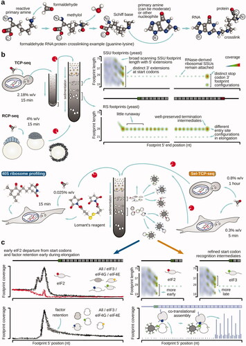
3,335
Views
2
CrossRef citations to date
0
Altmetric
Review Articles

Translation complex stabilization on messenger RNA and footprint profiling to study the RNA responses and dynamics of protein biosynthesis in the cells

Pages 261-304 | Received 23 Jun 2021, Accepted 11 Nov 2021, Published online: 01 Dec 2021

Figures & data

Figure 1. Overview of the protein biosynthesis pathway (translation). (a) The mRNA translation cycle of eukaryotic and prokaryotic cells, depicting the peptide initiation, elongation and termination, and ribosome recycling phases of the cycle. The focus is brought to the start codon location differences during the initiation phase. For more details about the initiation mechanisms, refer to the respective reviews (e.g. Shirokikh and Preiss Citation2018; Hinnebusch et al. Citation2016; Hinnebusch Citation2017; Hershey et al. Citation2019; Sonenberg and Hinnebusch Citation2009; Gualerzi and Pon Citation2015; Rodnina Citation2018). (b) Steps of translation elongation cycle as ribosome ratchets through the codons of an open reading frame of mRNA and polymerizes the polypeptide. The focus is brought to the events and intermediates that can be specifically targeted and/or can have an impact on the translation complex footprint features and distribution. A taxa-indifferent elongation factor designation is used, with EF1 referring to eEF1A in eukaryotes, aEF1A in archaea and EF-Tu in bacteria, and EF2 referring to eEF2 in eukaryotes, aEF2 in archaea and EF-G in bacteria. In the center of the cycle schematic, the respective aminoacyl (A), peptidyl (P), and exit (E) tRNA localization and functional sites of the SSU and LSU are highlighted (populated with tRNAs from the cycle schematic to provide an example), and the location of the peptidyl transferase center in the LSU (PTC) is schematized.

Figure 1. Overview of the protein biosynthesis pathway (translation). (a) The mRNA translation cycle of eukaryotic and prokaryotic cells, depicting the peptide initiation, elongation and termination, and ribosome recycling phases of the cycle. The focus is brought to the start codon location differences during the initiation phase. For more details about the initiation mechanisms, refer to the respective reviews (e.g. Shirokikh and Preiss Citation2018; Hinnebusch et al. Citation2016; Hinnebusch Citation2017; Hershey et al. Citation2019; Sonenberg and Hinnebusch Citation2009; Gualerzi and Pon Citation2015; Rodnina Citation2018). (b) Steps of translation elongation cycle as ribosome ratchets through the codons of an open reading frame of mRNA and polymerizes the polypeptide. The focus is brought to the events and intermediates that can be specifically targeted and/or can have an impact on the translation complex footprint features and distribution. A taxa-indifferent elongation factor designation is used, with EF1 referring to eEF1A in eukaryotes, aEF1A in archaea and EF-Tu in bacteria, and EF2 referring to eEF2 in eukaryotes, aEF2 in archaea and EF-G in bacteria. In the center of the cycle schematic, the respective aminoacyl (A), peptidyl (P), and exit (E) tRNA localization and functional sites of the SSU and LSU are highlighted (populated with tRNAs from the cycle schematic to provide an example), and the location of the peptidyl transferase center in the LSU (PTC) is schematized.

Figure 2. Schematic of popular approaches to study protein biosynthesis. (a) Approaches that provide a high-throughput surveillance of protein and RNA content or report on single-molecule activity of translating ribosomes and poly(ribo)somes. (b) Polysome profiling as one of the most broadly used methods to infer translational involvement across mRNA, including in a high-throughput format when employed together with microarrays or RNA-sequencing.

Figure 2. Schematic of popular approaches to study protein biosynthesis. (a) Approaches that provide a high-throughput surveillance of protein and RNA content or report on single-molecule activity of translating ribosomes and poly(ribo)somes. (b) Polysome profiling as one of the most broadly used methods to infer translational involvement across mRNA, including in a high-throughput format when employed together with microarrays or RNA-sequencing.

Figure 3. Schematic of nuclease probing methods to study translation. (a) A classical ribosome “footprinting” method using ribosome-protected ribonuclease (RNase)-resistant fragment identification specific to certain mRNAs of choice. (b) Translation (ribosome) footprint profiling, whereby the ribosome-protected RNase-resistant mRNA fragments are isolated based on their association with the ribosomes and then subjected to high-throughput RNA sequencing. Sedimentation-based isolation of the ribosome-associated mRNA fragments is exemplified, although other methods are possible (e.g. Reid et al. Citation2015). In the ribosome profiling, a high-throughput sequencing-derived coverage of a matching intact mRNA often serves as a coverage control and as a way of estimating the level of transnational involvement (“translation efficiency”) of the respective mRNAs (top right plot; gene-wize comparison for all genes). The relative footprint frequency over the respective ORF codons is often used to infer ribosome dwell time and the kinetic landscape of the ORF (bottom right plot; transcript-specific features).

Figure 3. Schematic of nuclease probing methods to study translation. (a) A classical ribosome “footprinting” method using ribosome-protected ribonuclease (RNase)-resistant fragment identification specific to certain mRNAs of choice. (b) Translation (ribosome) footprint profiling, whereby the ribosome-protected RNase-resistant mRNA fragments are isolated based on their association with the ribosomes and then subjected to high-throughput RNA sequencing. Sedimentation-based isolation of the ribosome-associated mRNA fragments is exemplified, although other methods are possible (e.g. Reid et al. Citation2015). In the ribosome profiling, a high-throughput sequencing-derived coverage of a matching intact mRNA often serves as a coverage control and as a way of estimating the level of transnational involvement (“translation efficiency”) of the respective mRNAs (top right plot; gene-wize comparison for all genes). The relative footprint frequency over the respective ORF codons is often used to infer ribosome dwell time and the kinetic landscape of the ORF (bottom right plot; transcript-specific features).

Figure 4. Examples of translation (ribosome) footprint profiling that use additional methods of targeted translation complex isolation, based on the presence of (left to right) specialized ribosomes, in trans interactions with the other translating ribosomes and their nascent peptides, tightly-packed nuclease-resistant stretches of ribosomes (e.g. most commonly, di- and tri-somes) and specific translation factors associated with the respective complexes.

Figure 4. Examples of translation (ribosome) footprint profiling that use additional methods of targeted translation complex isolation, based on the presence of (left to right) specialized ribosomes, in trans interactions with the other translating ribosomes and their nascent peptides, tightly-packed nuclease-resistant stretches of ribosomes (e.g. most commonly, di- and tri-somes) and specific translation factors associated with the respective complexes.

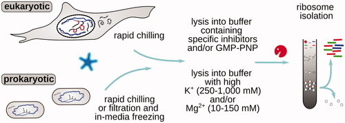
Table 1. Summary of the major approaches employed to arrest or stabilize translational complexes on mRNA for downstream footprinting and footprint profiling.

Figure 5. Popular specific inhibitors of translation elongation phase used to stabilize translational complexes in the footprint profiling research of eukaryotic cells. (a) Specific translation elongation inhibitors often used in translation footprint profiling research, their chemical structures and schematized mechanism of action (refer to the for more information on the elongation cycle context of the stabilized step). 3D (when available) and 2D structures of inhibitors were downloaded from PubChem (unless otherwise indicated). (b) Typical features of translation footprint patterns associated with the use of respective inhibitors (as indicated by arrows). Shown are schematized commonly observed variants of metagene or transcript-wise footprint distributions, aligned by footprint 5′ or 3′ ends across the sequence of a coding region of mRNA near the respective start (left, green) or stop (right, red) codons (see color version of this figure at www.tandfonline.com/ibmg).

Figure 5. Popular specific inhibitors of translation elongation phase used to stabilize translational complexes in the footprint profiling research of eukaryotic cells. (a) Specific translation elongation inhibitors often used in translation footprint profiling research, their chemical structures and schematized mechanism of action (refer to the Figure 1 for more information on the elongation cycle context of the stabilized step). 3D (when available) and 2D structures of inhibitors were downloaded from PubChem (unless otherwise indicated). (b) Typical features of translation footprint patterns associated with the use of respective inhibitors (as indicated by arrows). Shown are schematized commonly observed variants of metagene or transcript-wise footprint distributions, aligned by footprint 5′ or 3′ ends across the sequence of a coding region of mRNA near the respective start (left, green) or stop (right, red) codons (see color version of this figure at www.tandfonline.com/ibmg).

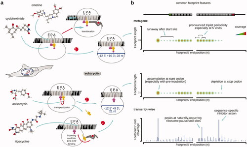
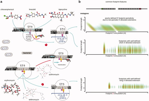
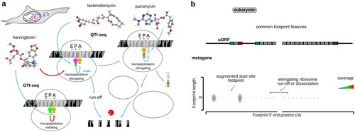
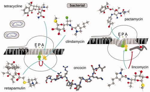
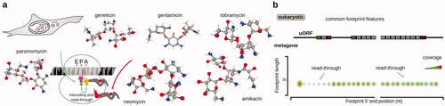
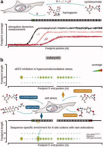
Table 2. Examples of antibiotics and inhibitors used in ribosomal footprinting research to study peptide elongation phase of protein biosynthesis.

Figure 6. Same as , but for translation elongation inhibitors commonly used in research of bacterial translation. Erythromycin structure (Fujii et al. Citation2013) was visualized using CCDC JSmol viewer.

Figure 6. Same as Figure 5, but for translation elongation inhibitors commonly used in research of bacterial translation. Erythromycin structure (Fujii et al. Citation2013) was visualized using CCDC JSmol viewer.

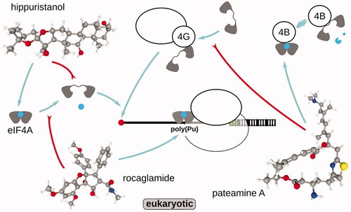
Table 3. Examples of antibiotics and inhibitors used in ribosomal footprinting research to study peptide initiation phase of protein biosynthesis.

Figure 7. Same as , but for translation inhibitors and their combinations commonly used to study eukaryotic translation initiation mechanisms.

Figure 7. Same as Figure 5, but for translation inhibitors and their combinations commonly used to study eukaryotic translation initiation mechanisms.

Figure 8. Small compounds targeting eukaryotic translation initiation factor and ATP-dependent single-stranded RNA binding protein (RNA helicase) eIF4A and its interactions with the substrates (mRNA, ATP) and other initiation factors and complexes, such as eIF4G:SSU and eIF4B. For more details about scanning and eIF4A action, refer to the specialized reviews (e.g. Shirokikh and Preiss Citation2018; Hinnebusch et al. Citation2016; Hinnebusch Citation2017; Naineni et al. Citation2020). Schematized mechanism of action of each compound is depicted. Note that in the majority of the observations, the footprint signal is found to be concentrated at or nearby the respective ORF start sites, and is observed in the footprints derived from the fraction of translational complexes containing complete ribosomes (as opposed to SSUs).

Figure 8. Small compounds targeting eukaryotic translation initiation factor and ATP-dependent single-stranded RNA binding protein (RNA helicase) eIF4A and its interactions with the substrates (mRNA, ATP) and other initiation factors and complexes, such as eIF4G:SSU and eIF4B. For more details about scanning and eIF4A action, refer to the specialized reviews (e.g. Shirokikh and Preiss Citation2018; Hinnebusch et al. Citation2016; Hinnebusch Citation2017; Naineni et al. Citation2020). Schematized mechanism of action of each compound is depicted. Note that in the majority of the observations, the footprint signal is found to be concentrated at or nearby the respective ORF start sites, and is observed in the footprints derived from the fraction of translational complexes containing complete ribosomes (as opposed to SSUs).

Figure 9. Same as , but for translation inhibitors commonly used to study bacterial translation initiation mechanisms. Oncocin Onc112 PDB structure 4ZER was visualized using PDB Mol viewer (Seefeldt et al. Citation2015).

Figure 9. Same as Figure 7, but for translation inhibitors commonly used to study bacterial translation initiation mechanisms. Oncocin Onc112 PDB structure 4ZER was visualized using PDB Mol viewer (Seefeldt et al. Citation2015).

Figure 10. Specific translation termination inhibitors often used in translation footprint profiling research. (a) Mechanism of action and the stabilized state of the ribosome characteristic to the exemplified compounds. Note that the currently used translation termination inhibitors are as such inducers of read-through and thus, unlike inhibitors used to study the other phases of translation, in fact de-stabilize translation termination complexes. The shown compounds have a broad specificity across taxa and are used in both, eukaryotic and bacterial research. (b) Typical meatgene features of translation footprint patterns associated with the use of respective inhibitors (for designations, refer to ) using an eukaryotic system as example.

Figure 10. Specific translation termination inhibitors often used in translation footprint profiling research. (a) Mechanism of action and the stabilized state of the ribosome characteristic to the exemplified compounds. Note that the currently used translation termination inhibitors are as such inducers of read-through and thus, unlike inhibitors used to study the other phases of translation, in fact de-stabilize translation termination complexes. The shown compounds have a broad specificity across taxa and are used in both, eukaryotic and bacterial research. (b) Typical meatgene features of translation footprint patterns associated with the use of respective inhibitors (for designations, refer to Figure 5(b)) using an eukaryotic system as example.

Table 4. Examples of antibiotics and inhibitors used in ribosomal footprinting research to study peptide termination and ribosomal recycling phases of protein biosynthesis.

Figure 11. Combinations of specific translation inhibitors and their effects to study the dynamics of translation elongation and translation complex identity, as well as codon specificity, of the stalled intermediates in eukaryotic systems. (a) Cycloheximide chase following harringtonin translation initiation arrest to infer the rate of ribosomal progression along mRNA ORFs. Note that the translation initiation is stopped upon the completion of the start codon recognition and the assembly of the elongation-capable ribosome at the respective start sites. (b) Use of tigecycline-cycloheximide and anisomycin-cycloheximide combinations to arrest ribosomes with differently configured A-sites, populated (left, bottom; blocked transpeptidation) and non-populated (right, top; blocked aminoacyl-tRNA accommodation) with the incoming aminoacyl-tRNA:eEF1A:GTP, respectively. Shorter footprints (left, bottom) in this case are reflective of the delays in attracting the respective aminoacyl-tRNA, whereas longer footprints (right, top) are reflective of eEF2 deficiency (see text for more details).

Figure 11. Combinations of specific translation inhibitors and their effects to study the dynamics of translation elongation and translation complex identity, as well as codon specificity, of the stalled intermediates in eukaryotic systems. (a) Cycloheximide chase following harringtonin translation initiation arrest to infer the rate of ribosomal progression along mRNA ORFs. Note that the translation initiation is stopped upon the completion of the start codon recognition and the assembly of the elongation-capable ribosome at the respective start sites. (b) Use of tigecycline-cycloheximide and anisomycin-cycloheximide combinations to arrest ribosomes with differently configured A-sites, populated (left, bottom; blocked transpeptidation) and non-populated (right, top; blocked aminoacyl-tRNA accommodation) with the incoming aminoacyl-tRNA:eEF1A:GTP, respectively. Shorter footprints (left, bottom) in this case are reflective of the delays in attracting the respective aminoacyl-tRNA, whereas longer footprints (right, top) are reflective of eEF2 deficiency (see text for more details).

Figure 12. Commonly accepted inhibitor-free approaches to preserve translational complexes on mRNA.

Figure 12. Commonly accepted inhibitor-free approaches to preserve translational complexes on mRNA.

Figure 13. Overview of translation complex footprinting methods based on in vivo stabilization of translation intermediates with covalent crosslinking. (a) Example of a possible formaldehyde-mediated RNA-protein crosslink formation. (b) Schematized TCP-seq, RCP-seq, 40S ribosome profiling and Sel-TCP-seq methods (see text for more details). Characteristic footprint patterns and features for both, SSU- and ribosome-derived footprints are shown, covering the entire translation cycle on mRNA. (c) Factor-selective methods using covalent stabilization allow to obtain additional insight into translation factor involvement along the translation cycle phases (left), and refine start selection mechanisms and dynamics, as well as detect and monitor co-translational complex assembly (right). Designations and structures as in .

Figure 13. Overview of translation complex footprinting methods based on in vivo stabilization of translation intermediates with covalent crosslinking. (a) Example of a possible formaldehyde-mediated RNA-protein crosslink formation. (b) Schematized TCP-seq, RCP-seq, 40S ribosome profiling and Sel-TCP-seq methods (see text for more details). Characteristic footprint patterns and features for both, SSU- and ribosome-derived footprints are shown, covering the entire translation cycle on mRNA. (c) Factor-selective methods using covalent stabilization allow to obtain additional insight into translation factor involvement along the translation cycle phases (left), and refine start selection mechanisms and dynamics, as well as detect and monitor co-translational complex assembly (right). Designations and structures as in Figure 5.

Figure 14. Formaldehyde-based covalent stabilization of translational complexes is a versatile approach compatible with rapid protein biosynthesis dynamics. (a) Estimates of formaldehyde vs. cycloheximide characteristic diffusion times to a given % of their initial concentration (blue line demarks 90%) over a typical barrier (5 µm for attached cell thickness or 50 µm for a group of cells or tissue fragment). Diffusion coefficients in water and a 2-term Taylor series approximation of the Fick’s law were used; calculations and plotting was done in GeoGebra (https://www.geogebra.org). (b,c) Mapping of HEK 293 formaldehyde-fixed TCP-seq-style footprints demonstrating the presence of footprints from nuclease-resistant disome fraction (b), specific signals from mitochondrial SSU and ribosomes in the respective fractions, and mitochondrial gene coverage in all, SSU, ribosome and disome fractions (c). These results suggest universal and taxonomically indifferent applicability of the formaldehyde-based covalent stabilization of translational complexes (see color version of this figure at www.tandfonline.com/ibmg).

Figure 14. Formaldehyde-based covalent stabilization of translational complexes is a versatile approach compatible with rapid protein biosynthesis dynamics. (a) Estimates of formaldehyde vs. cycloheximide characteristic diffusion times to a given % of their initial concentration (blue line demarks 90%) over a typical barrier (5 µm for attached cell thickness or 50 µm for a group of cells or tissue fragment). Diffusion coefficients in water and a 2-term Taylor series approximation of the Fick’s law were used; calculations and plotting was done in GeoGebra (https://www.geogebra.org). (b,c) Mapping of HEK 293 formaldehyde-fixed TCP-seq-style footprints demonstrating the presence of footprints from nuclease-resistant disome fraction (b), specific signals from mitochondrial SSU and ribosomes in the respective fractions, and mitochondrial gene coverage in all, SSU, ribosome and disome fractions (c). These results suggest universal and taxonomically indifferent applicability of the formaldehyde-based covalent stabilization of translational complexes (see color version of this figure at www.tandfonline.com/ibmg).