Figures & data
Figure 1. Application strategies for nanoparticle-mediated drug delivery in pregnancy. Three therapeutic scenarios are depicted: (A) treatment of maternal conditions without fetoplacental exposure; (B) treatment of placental conditions without maternal or fetal exposure; (C) treatment of fetal conditions without maternal or placental exposure.
Table 1. Representative studies exploring the application of nanotechnology for placenta-originated disease therapy.
Figure 2. Diagram of the physiological structure of the placenta. (A) Modified spiral arteries enable sufficient perfusion of the placenta with maternal blood that bathes the intervillous space and makes direct contact with the SCT. (B) SCT is a master regulator of placental translocation. (C) Fetal blood enters the placenta via the umbilical artery (blue) and flows into the capillaries in the placental villi before returning to the fetus via the umbilical vein (red). (SCT: syncytiotrophoblasts, VCT: villous cytotrophoblast, TGC: trophoblast giant cells, HB: Hofbauer cells, FB: fibroblasts, CCC: cytotrophoblast cell column, DC: decidual cells, iEVT: interstitial extravillous trophoblast, enEVT: endovascular extravillous trophoblast, egEVT: endoglandular extravillous trophoblast, ULE: uterine luminal epithelium, FEC: fetal endothelial cell). Image from reference (Arumugasaamy et al., Citation2020) cited with permission. Copyright © 2020 Elsevier B.V.
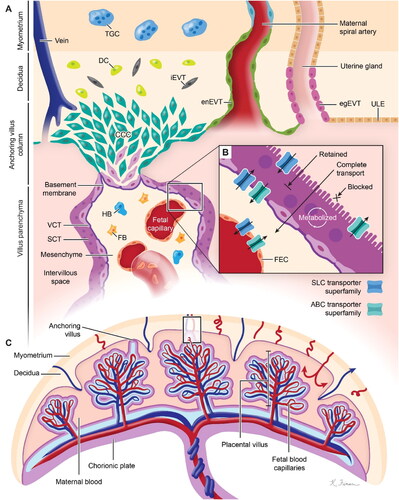
Figure 3. Transport of nanodrugs in the placenta. Trophoblast uptake through the paracellular pathway (A), endocytosis (B), and transporter-mediated pathway (C). Trophoblast exocytosis of nanoplatforms (D).
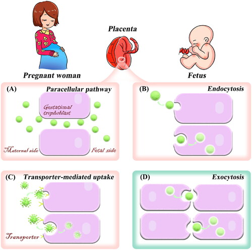
Figure 4. Pregnant mice injected intravenously with Cy 5 solution or Cy 5-loaded liposomes of different sizes were processed and analyzed after 2 and 8 h. (A) Fluorescent sections containing Cy 5 in the placenta and fetus of pregnant mice. (B) Cy 5 fluorescence intensity in placenta and fetus of pregnant mice. (C) Quantitative analysis of Cy 5 content in the placenta and fetal tissues of pregnant mice by liquid chromatography-mass spectrometry. Image from reference (Tang et al., Citation2022) cited with permission. Copyright © 2022 Elsevier B.V.
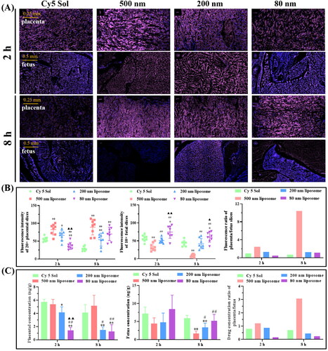
Table 2. Characteristics of nanoplatforms that would be beneficial in treating pregnancy complications.
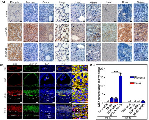
Figure 5. (A) Analysis of sections of different tissue blocks incubated with anti-C4S (2B6) and plCSA-BP revealed that plCSA-BP specifically binds to placental tissue. (B) plCSA-BP specifically binds to trophoblast at different gestational stages in mice. Trophoblast cells (CK8, red), biotin-plCSA-BP (green), and nuclei (DAPI, blue). Dec: decidua; Em: embryo; Jz: junctional zone; Lab: labyrinth. (C) Quantitative analysis of MTX concentrations in placenta and fetuses by HPLC. nd: not detected. MNPs: Lipid-polymer nanoparticles loaded with MTX; SCR-MNPs: The scrambled peptide-conjugated nanoparticles loaded with MTX; plCSA-MNPs: plCSA-BP-conjugated nanoparticles loaded with MTX. Image from reference (Zhang et al., Citation2018b) cited with permission. Copyright © Ivyspring International Publisher.