Figures & data
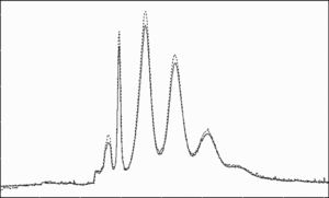
Figure 1 The electropherogram of SC-PEG-bHb (the molar ratio of hemoglobin to SC-PEG was 1 : 8) by capillary zone electrophoresis.
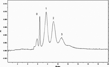
Figure 2 The SE-HPLC profile of disassociated SC-PEG-bHb. (mobile: 5 mM sodium phosphate, 150 mM NaCl, 1 M MgCl2, pH 7.0; UV : 280 nm; Flow rate: 1 ml/min).
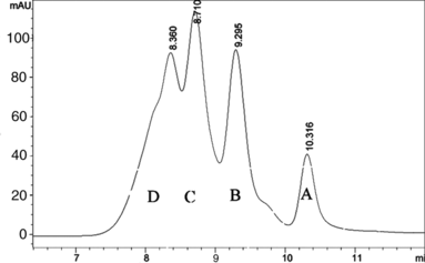
Figure 3 SDS-PAGE analysis of each fraction of SE-HPLC stained with Coomassie blue. (Lane1, 2, 3, and 4 represent the fractions A, B, C, and D in Fig. 2, and Lane 5 is the molecular mark).
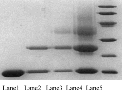
Figure 4 The electropherogram of fractions A, B, C, and D of SE-HPLC by capillary zone electrophoresis.
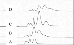
Figure 5 Electropherogram of PEG-bHb produced by different reaction ratios of hemoglobin to PEG (A,1:8; B,1:10; C,1:12).
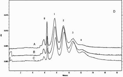
Figure 6 The electropherogram of SC-PEG-bHb before (solid line) and after (dot line) incubating with 10 mM hydroxylamine (molar ratio 1:12).
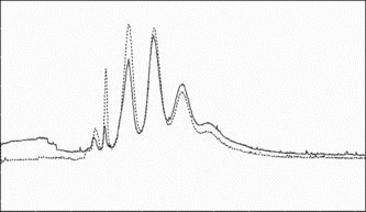
Figure 7 The electropherogram of SPA-PEG-bHb before (solid line) and after (dot line) incubating with 10 mM hydroxylamine (molar ratio 1:12).