483
Views
11
CrossRef citations to date
0
Altmetric
Original

Thiolation Mediated Pegylation Platform to Generate Functional Universal Red Blood Cells

, &
Pages 107-118 | Published online: 24 Aug 2009

Figures & data

Figure 1 Schematic representation of Protocols used for PEGylation of RBC. Scheme 1: Iminothiolane-thiolation mediated PEGylation. Iminothiolane reacts with amino groups generating thiol groups in situ. These thiols are modified by maleimide-PEG (Mal-Phe-PEG). Scheme 2: DTSSP-thiolation mediated PEGylation. DTSSP is a cross-linking reagent and uses acylation chemistry to react with amino groups. On treatment with reducing agents, the S–S bond generates free thiols that can be modified by Mal-Phe-PEG. Scheme 3: Acylation chemistry mediated PEGylation. Succinimidyl propionate-PEG (SPA-PEG) acylates amino groups of proteins. In schemes 1 and 2 an extension arm is added on protein amino groups for PEGylation and in scheme 3 PEGylation of proteins is carried out without adding extension arm.
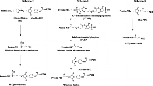
Figure 1 Schematic representation of Protocols used for PEGylation of RBC. Scheme 1: Iminothiolane-thiolation mediated PEGylation. Iminothiolane reacts with amino groups generating thiol groups in situ. These thiols are modified by maleimide-PEG (Mal-Phe-PEG). Scheme 2: DTSSP-thiolation mediated PEGylation. DTSSP is a cross-linking reagent and uses acylation chemistry to react with amino groups. On treatment with reducing agents, the S–S bond generates free thiols that can be modified by Mal-Phe-PEG. Scheme 3: Acylation chemistry mediated PEGylation. Succinimidyl propionate-PEG (SPA-PEG) acylates amino groups of proteins. In schemes 1 and 2 an extension arm is added on protein amino groups for PEGylation and in scheme 3 PEGylation of proteins is carried out without adding extension arm.

Table 1 Functional and serological properties of PEGylated RBCs

Figure 2 Microscopic evaluation of agglutination of PEGylated RBCs in the presence of anti-D. RBCs are PEGylated by (A) imnothiolane-thiolation mediated PEGylation using Mal-Phe-PEG-5000, (B) DTSSP-based-thiolation mediated PEGylation using Mal-Phe-PEG-5000, and (C) Acylation chemistry mediated PEGylation using SPA-PEG-5000.

Figure 2 Microscopic evaluation of agglutination of PEGylated RBCs in the presence of anti-D. RBCs are PEGylated by (A) imnothiolane-thiolation mediated PEGylation using Mal-Phe-PEG-5000, (B) DTSSP-based-thiolation mediated PEGylation using Mal-Phe-PEG-5000, and (C) Acylation chemistry mediated PEGylation using SPA-PEG-5000.

Figure 3 Microscopic evaluation of agglutination of PEGylated RBCs in the presence of anti-A (A, C & E) and anti-D (B, D and F). RBCs are PEGylated by (A & B) imnothiolane-thiolation mediated PEGylation using Mal-Phe-PEG-5000 in step-1 and Mal-Phe-PEG-20000 in step-2, (C & D) DTSSP-thiolation mediated PEGylation using Mal-Phe-PEG-5000 in step-1 and Mal-Phe-PEG-20000 in step-2 (E & F) acylation chemistry mediated PEGylation using SPA-PEG-5000 in step-1 and SPA-PEG-20000 in step-2.
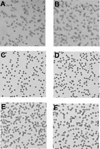
Figure 3 Microscopic evaluation of agglutination of PEGylated RBCs in the presence of anti-A (A, C & E) and anti-D (B, D and F). RBCs are PEGylated by (A & B) imnothiolane-thiolation mediated PEGylation using Mal-Phe-PEG-5000 in step-1 and Mal-Phe-PEG-20000 in step-2, (C & D) DTSSP-thiolation mediated PEGylation using Mal-Phe-PEG-5000 in step-1 and Mal-Phe-PEG-20000 in step-2 (E & F) acylation chemistry mediated PEGylation using SPA-PEG-5000 in step-1 and SPA-PEG-20000 in step-2.

Reprints and Corporate Permissions

Please note: Selecting permissions does not provide access to the full text of the article, please see our help page How do I view content?

To request a reprint or corporate permissions for this article, please click on the relevant link below:

Academic Permissions

Please note: Selecting permissions does not provide access to the full text of the article, please see our help page How do I view content?

Obtain permissions instantly via Rightslink by clicking on the button below:

If you are unable to obtain permissions via Rightslink, please complete and submit this Permissions form. For more information, please visit our Permissions help page.