Figures & data
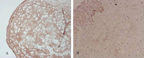
Figure 1. The results of Col II specific staining (×200). A: Col II specific staining of induced cells; B: Col II specific staining of rat chondrocytes; C: Col II specific staining of rat BMSC.
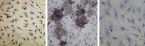
Figure 2. Detection of collagen type II and aggrecan mRNA expression by RT-PCR. 1: RNA marker; 2: Col II expression of BMSCs without induction; 3: Col II expression of induced cells; 4: Col II expression of chontrocytes; 5: aggrecan expression of induced cells; 6: aggrecan expression of chontrocytes; 7: aggrecan expression of BMSCs without induction.
Figure 3. PGA/PLA scaffolds.
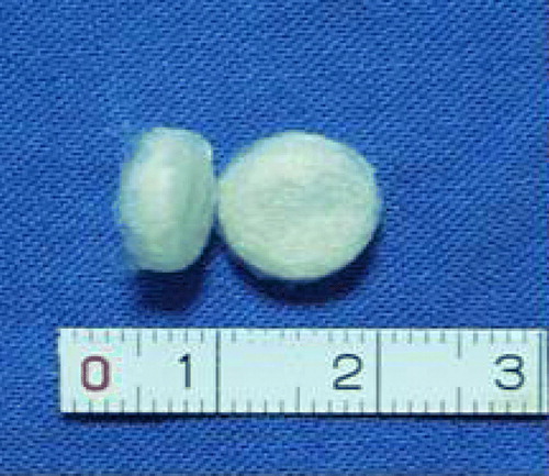
Figure 4. Cell attachment on scaffold.
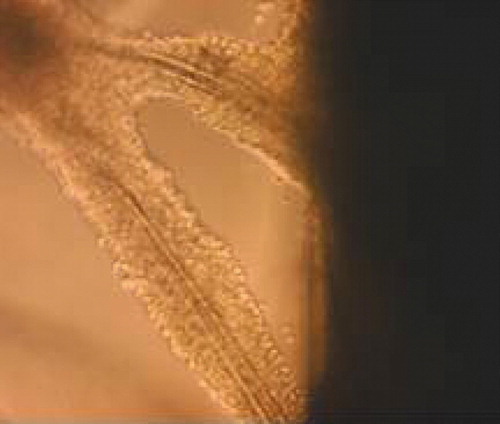
Figure 5. Gross view of specimens in all groups cultured in vitro for 8 weeks. A: Cells and scaffold (24h); B: co-culture group; C: Chondrocyte group; D: BMSC group; E: Low chondrocytes group.
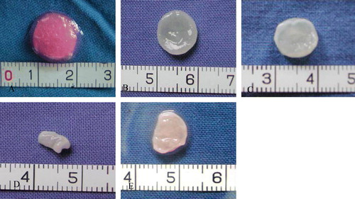
Figure 6. Histology of specimens in all groups cultured in vitro for 8 weeks. A: co-culture group; B: Chondrocyte group; C: BMSC group; D: Low chondrocytes group.
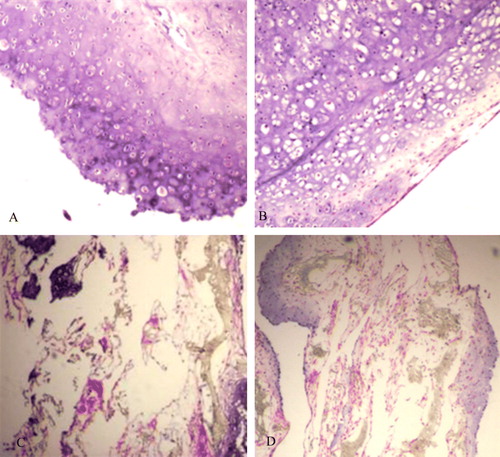
Figure 7. Massion strain of some specimens in vitro for 8 weeks. A: co-culture group; B: Chondrocyte group.
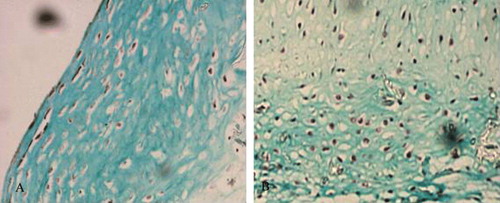
Figure 8. Safranin O strain of some specimens in vitro for 8 weeks. A: co-culture group; B: Chondrocyte group.
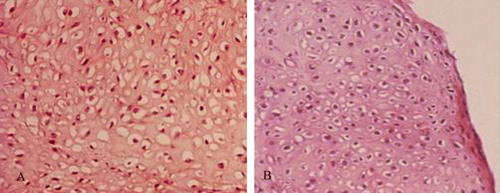
Figure 9. Strain of some specimens in vitro for 8 weeks. A: co-culture group; B: Chondrocyte group.
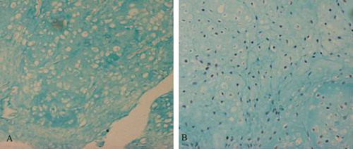
Figure 10. Collagen type II immunohistochemistry of some specimens cultured in vitro for 8 weeks. A: co-culture group; B: Chondrocyte group.