Figures & data
Figure 1. Floripatella rousseaui Yochelson, Citation1988, USNM PAL 410165, holotype, internal mould, Middle Ordovician, Darriwilian, Kanosh Shale, Millard County, Utah. A. lateral view oblique to the plane of symmetry B where large arrows locates intersection of a radial crack with the muscle scar in B and D. B. dorsal view with arrow pv locating the putative pallial vessel impression discussed by Lindberg (Citation2009, fig. 6). C. detail of muscle scar showing median ridge (mr) and one discrete scar (ds). D. dorsal view, as B but with alternative lighting, showing radial muscle fibres (rmf) and pericardium depression (pd). E. detail of margin with pallial vessel. Scale bars: 3 mm, C,E; 5 mm, A,B,D. Photographs prepared from negatives supplied by E.L. Yochelson.
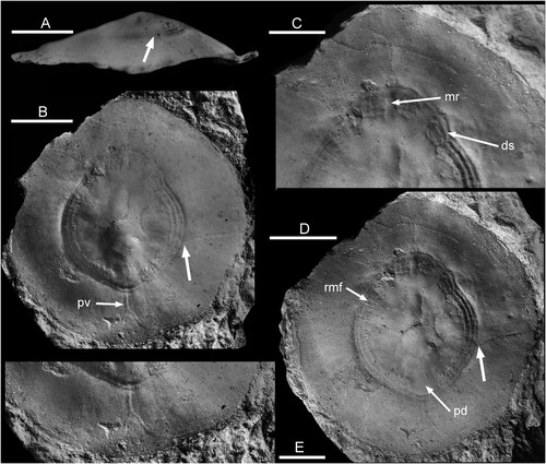
Figure 2. Pollicina Koken in Holzapfel, Citation1895, Ordovician, Darriwilian, Kunda Stage. A–E. Pollicina crassitesta Koken, Citation1897, internal moulds. A,B. ELM G1:2919, sub-apical and lateral views with muscle attachment scar, Tallinn. C,D. ELM g1:2323, oblique views of supra-apical surface with muscle attachment scar, Tallinn. E. CNIGRM 15600, lateral view with muscle attachment scar, Laaksberg ( = Lasnamägi), Estonia. F. Pollicina corniculum (Eichwald, Citation1860), CNIGRM 15702, lateral view showing comarginal ornamentation on shell exterior, Pulkowa (=Pulkovo), St. Petersburg, Russia. Scale bars: 2 mm, C; 4 mm, A,B,D,E; 5 mm, F.
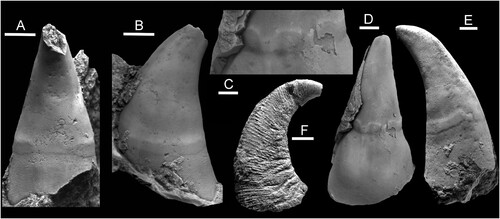
Figure 3. Eesticonus aariensis n. gen. n. sp., TUG 1787-21, holotype, internal mould with broken apex showing muscle attachment scars. Ordovician, Darriwilian Series, Kunda Stage, Aari Quarry Noonu Parish, Haljala, northern Estonia. A. lateral view. B. supra-apical surface. C. oblique view of sub-apical surface showing trace of shell laminae (tl). D. dorsal view, supra-apical surface to left. E. oblique view of supra-apical surface, with radially ornamented shell fragment (arrow). F. tubercles on the internal mould representing pits in the dissolved shell, located by arrow F in G. G. oblique view of supra-apical surface with raised comarginal band (arrow cb) that may represent an endolith burrow; position of Fig. F indicated by arrow. H. oblique view of supra-apical surface showing medial (ma) and lateral (la) angulations; es indicates the extension of the muscle scar band towards the median plane. Scale bars: 2 mm, F; 6 mm, A–E,G,H.
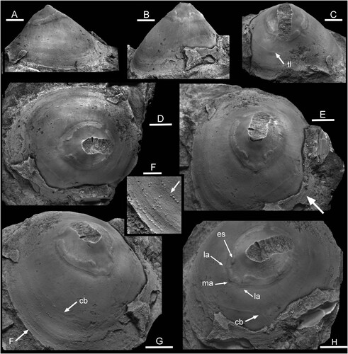
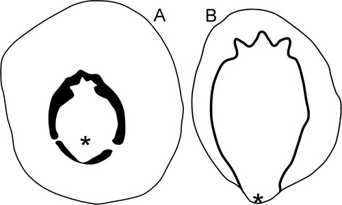
Figure 4. Drawings of the muscle attachment scars on internal moulds of the holotype of Eesticonus aariensis (A) and a paratype (NM L 5903) of Archinacellina modesta (B), the latter based on Peel & Horný (Citation1999, fig. 10A). Specimens are oriented as gastropods, with the anterior uppermost. Asteriscs locate position of shell apex.