
Figures & data
Figure 1. Photography of the POCUS Butterfly-iQ device connected to the smartphone used in this study. OB 1/GYN was the chosen preset.
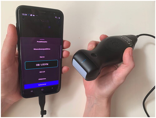
Figure 2. Illustration of the IUD-myometrium distance used in the current study (dashed line in blue).
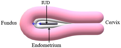
Table 1. Baseline characteristics of subjects.
Table 2. Detailed IUD position found by abdominal POCUS and conventional transvaginal US.
Table 3. Diagnostic performance of abdominal POCUS in predicting IUD malposition.
Table 4. Agreement between abdominal POCUS and conventional transvaginal US in detecting IUD position.
Figure 3. Ultrasonographic images. A - copper IUD entirely within the uterine cavity in a retroverted uterus by both POCUS (A1) and conventional US (A2); B - hormonal IUD entirely within the uterine cavity in an anteverted uterus by both POCUS (B1) and conventional US (B2). The top and the distal end of the IUDs are indicated by yellow arrows.
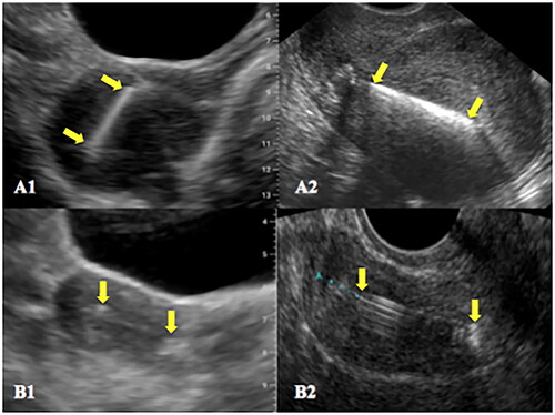
Figure 4. Ultrasonographic images. C - copper IUD entirely within the cervical canal by both POCUS (C1) and conventional US (C2); D - copper IUD embedded in the myometrium by both POCUS (D1) and conventional US (D2). The top and the distal end of the IUDs are indicated by yellow arrows. Asterisks indicate the fundal region of the endometrial cavity.
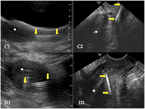
Figure 5. Ultrasonographic images. E - hormonal IUD identified as entirely within the uterine cavity by POCUS (E1) and the same IUD with a small portion within the cervical canal identified by conventional US (E2); F - a small part of a copper IUD apparently within the cervical canal found by POCUS (F1) and the same IUD entirely within the uterine cavity by conventional US (F2). The top and the distal end of the IUDs are indicated by yellow arrows. In the image F1, the proximal portion of the string was probably mistaken for the end of the IUD.
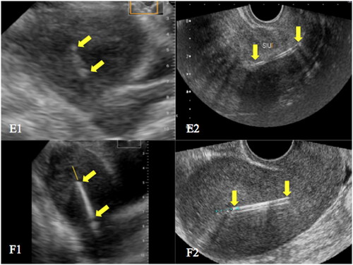
Figure 6. The concordance plot of IUD-myometrium distance between POCUS and conventional US.
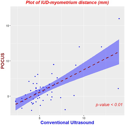
Table 5. Case-by-case differences in IUD-myometrium distance and subjective impression for low-lying IUDs found by POCUS and/or conventional US.
Table 6. Clinical and ultrasound features that may affect the agreement between abdominal POCUS and conventional transvaginal US.