Figures & data
Figure 1. Acanthosis nigricans. Gray-brown to black lesions, rough, thickened plaques and prominent skin lines.
Table I. Effects of testosterone in skin diseases
Figure 2. Skin in hypogonadism. The skin is thin and smooth; wrinkles are notably small and crinkling.
Figure 3. Androgenic alopecia. In an advanced stage, the bald skin of the scalp is surrounded by a chaplet.
Figure 4. The typical flush, the clinical hallmark of the carcinoid syndrome, begins suddenly and lasts 20 to 30 seconds. It primarily involves the face, neck, and upper chest. (Courtesy of Prof. Dr Arnold, Marburg, Germany)
Table II. Clinical signs in Cushing's disease
Figure 5. Topical overdosage of corticosteroids. Following long-term application, development of telangiectasias and a rosacea-like dermatitis may occur, particularly on the face.
Figure 6. The diabetic foot. Chronic inflammations of the skin with defects and subsequent ulcers, showing recurrent infections.
Table III. Diabetic foot care
Figure 7. Necrobiosis lipoidica: Yellowish-brown lesions with red margin. The epidermis is thin and shows little scaling; ulceration is common.
Figure 8. Gynecomastia. Large pending breast with significant submammary fold in a patient with Klinefelter's syndrome.
Figure 9. Necrolytic migratory erythema in glucagonoma. Bronze-colored, indurated areas with blistering, crusting, and scaling at the borders.
Figure 10. Acromegaly. Thickening, pasty consistence, thickening of skin folds, thickening of lips and eyelids, hypertrichosis, bushy eyebrows, growth of bones, enlargment of the ears, protrusion of zygoma, mandible and chin.
Table IV. Clinical signs of acromegaly.
Figure 11. Myxedema. Pasty and voluminous skin; non-impressible not position-dependent edema.
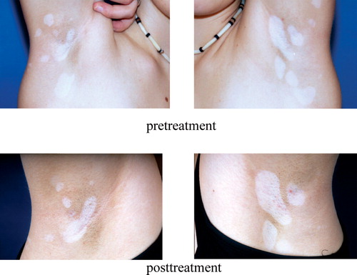
Figure 12. Vitiligo. Local loss of skin pigmentation due to the malfunction or loss of melanocytes. After three months' treatment with tacrolimus, the size of lesions is decreased.