
Figures & data
Figure 1. Pathological results of patient after TURBT. (A) HE staining (×400) showed the pathological type of high-grade uroepithelial carcinoma; (B) IHC (×400) showed positive AE1/AE3 expression; (C) IHC (×400) showed positive Ki67 expression (index 80%); (D) IHC (×400) showed positive P40 expression; (E) IHC (×400) showed positive P53 expression, 90%+; (F) IHC (×400) showed shows positive P63 expression.
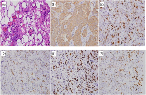
Figure 2. Partial imaging examinations after the patient developed metastatic symptoms. (A) whole-body bone tomography showed active bone salt metabolism in lumbar 1–4 vertebrae, mostly considering bone metastases; (B) chest CT showed multiple enlarged lymph nodes in the left side of the neck, considering possible metastases; (C,D) whole-abdomen CT showed multiple retroperitoneal lymph node metastases; (E,F) whole-services CT showed multiple pelvic lymph node metastases.
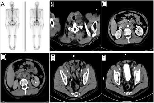
Figure 3. Cervical lymph node biopsy results. (A) HE staining (×400) showed indicates migratory cell carcinoma on cervical lymph node puncture; (B) IHC (×400) showed positive CAm5.2 expression; (C) IHC (×400) showed positive CK-P expression; (D) IHC (×400) showed positive CK8/18 expression; (E) IHC (×400) showed positive HER2 expression (3+); (F) IHC (×400) showed positive Ki67 expression; (G) IHC (×400) showed positive P40 expression; (H) IHC (×400) showed positive P63 expression; (I) IHC (×400) showed positive Uroplakin expression.
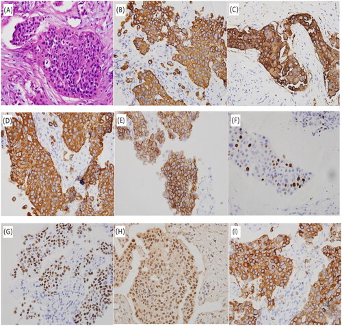
Figure 4. Partial imaging examinations after regular chemotherapy. (A) whole-body bone tomography showed that the bone salt metabolism of the lumbar 1–4 vertebrae was lower than before, the density was higher than before, and the range of osteogenesis was expanded. (B) Magnetic resonance imaging of the neck showed that multiple enlarged lymph nodes were seen in the neck area IV and the left clavicle fossa, both D1WI and T2WI showed high signal, abnormal lymph nodes were considered. (C,D) Whole abdominal CT showed abnormal abdominal lymph nodes disappeared. (E,F) Whole abdominal CT showed abnormal pelvic lymph node disappeared.
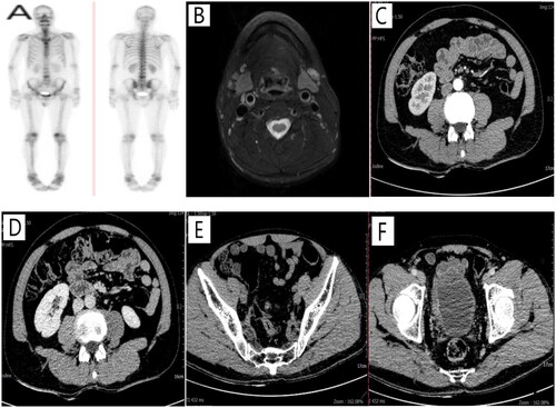
Table 1. cases of bladder cancer presenting with supradiastinal lymph nodes metastasis after surgery.