
Figures & data
Figure 1. MRI: prostate size was about 57 mm × 50 mm × 54 mm, increased prostatic transitional zone, and surrounding of prostatic duct indicate bar isointense T1 and short T2; posterior of peripheral zone indicate patchy isointense T1 and short T2.
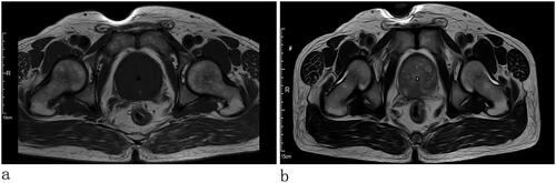
Figure 2. Chest CT: no tumor in the lungs and pleura (a) transverse view and (b) coronal view). (c) Whole body bone scan: no bone metastasis.
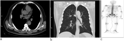
Figure 3. PET/CT: prostate protruding into the bladder was enlarged, with abnormal increases in glucose metabolism of the local lesion, considered to be a malignant tumor, and hypermetabolic pelvic lymph nodes, which should be considered for metastasis.
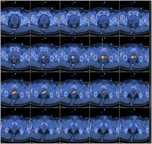
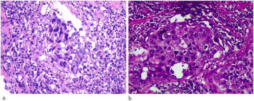
Figure 4. (× 400 HE) The tumor cells exhibited Umbrella or spindle-shaped and were diffusely nests and clumps, cleft formation in partial cells regions, a necrotic area in some areas, with abundant clear cytoplasm, conspicuous nucleoli, mark nuclear atypia, and numerous karyokinesis.
Data availability statement
All data generated or analyzed during this study are included in this published article.