Figures & data
Table 1. The effects of the ethyl acetate root extract D. tripetala. on packed cell volume (PCV %) of mice.
Table 2. The effects of ethyl acetate root extract of D. tripetala. on red blood cell count in mice (no./µl × 106).
Figure 1 The effect of the ethyl acetate root extract of D. tripetala. on total white blood cell count in mice.
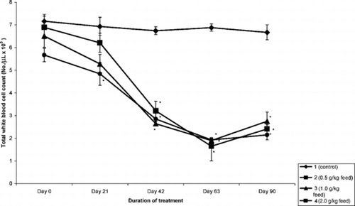
Figure 2 The effect of the ethyl acetate root extract of D. tripetala. on lymphocyte count in mice.
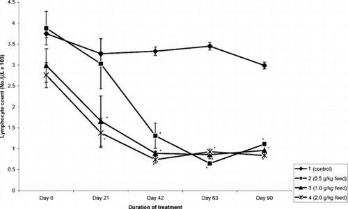
Figure 3 The effect of the ethyl acetate root extract of D. tripetala. on neutrophil count in mice.
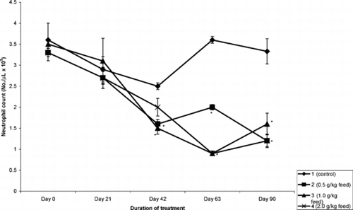
Table 3. The relative organ weights (mean ± SEM) of mice treated with ethyl acetate extract of D. tripetala. in mice.
Figure 4 Liver section of untreated (group 1) mouse collected on day 42 of study showing hepatic artery (R) and hepatic portal vein (V) in the portal area. Hepatocytes with vesicular nuclei in cords (arrowheads). H&E stain, ×400.
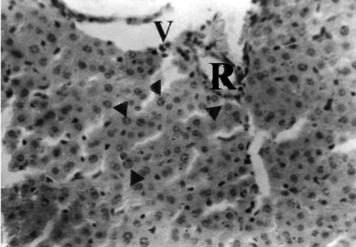
Figure 5 Liver section of mouse in group 3 (1.0 g/kg feed) collected on day 21 of treatment showing hepatocytes undergoing necrosis with pyknotic nuclei (arrowheads) in the portal area with hepatic artery (R). H&E stain, ×400.
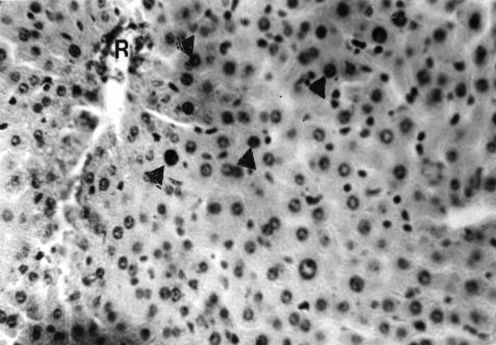
Figure 6 Liver section of mouse in group 3 (1.0 g/kg feed) collected after 63 days of treatment showing widespread vacuolation of hepatocytes and pyknosis of nuclei (arrowhead). H&E stain, ×400.
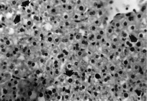
Figure 7 Liver section of mouse in group 4 (2.0 g/kg feed) collected after 42 days of treatment showing widespread necrosis of hepatocytes and many mononuclear leukocytes (arrowheads) around the biliary ductules (B) and the entire portal area. H&E stain, ×400.
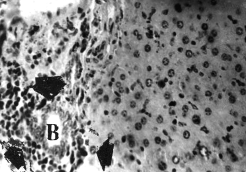
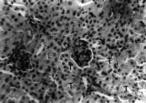
Figure 8 Kidney section of untreated (group 1) mouse collected on day 42 of study showing renal corpuscles (arrowheads) and normal epithelial cells lining the tubules (T). H&E stain, ×400.
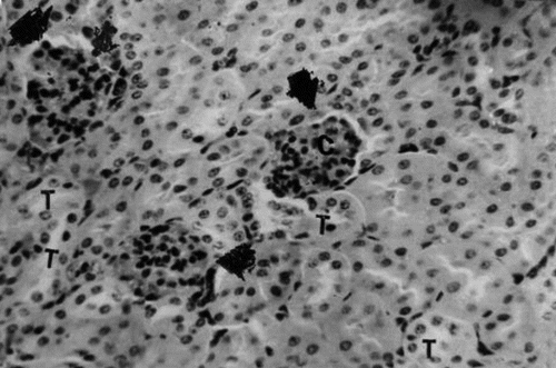
Figure 9 Kidney section of group 3 (1.0 g/kg feed) mouse collected on day 21 of treatment showing degeneration and necrosis of epithelial cells lining the tubules (T). H&E stain, ×400.
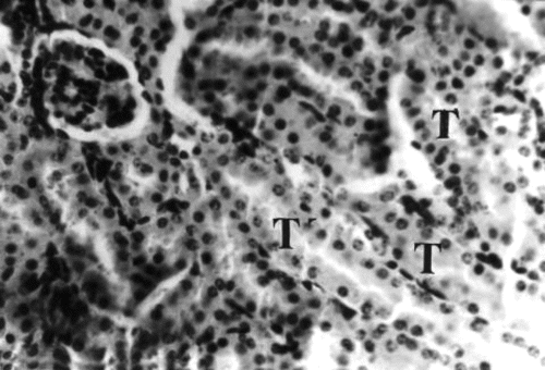
Figure 10 Kidney section of group 4 (2.0 g/kg) mouse collected on day 21 of treatment showing widespread pyknosis of tubular (T) epithelial cells and mononuclear leukocytes infiltration of interstices and perivascular spaces, H&E stain, ×400.