Figures & data
Figure 1 Chemical structure of salidroside.
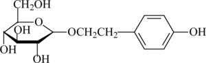
Figure 2 Separation and purification of salidroside from extracts of Rhodiola rosea..

Figure 3 Effects of Na2S2O4-induced cytotoxicity on PC12 cells. Confluent cells in 96-well plates were treated with different concentrations in the presence of Na2S2O4 and then changed to normal medium for 2 h. Cell viability was determined by the MTT assay, and the data were expressed as X ± SD, n = 3. *p < 0.05, **p < 0.01, and ***p < 0.001 in comparison with normal cells.
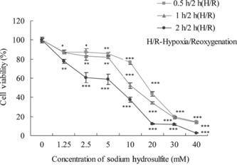
Figure 4 Toxicologic effects of salidroside on PC12 cells. Confluent PC12 cells, exposed to different concentrations of salidroside, were preincubated in 96-well plates for 24 h. Data were expressed as X ± SD, n = 3.
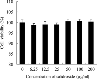
Figure 5 The stability of salidroside in RPMI 1640 medium. Salidroside (100 µg/mL) was dissolved in RPMI 1640 medium and then incubated at 37°C in a humidified atmosphere of 95% air and 5% CO2. The solution was centrifuged at 10,000 rpm for 15 min at 4°C, and then the supernatant was filtrated through an 0.45-µm membrane. The supernatant was assayed by HPLC.
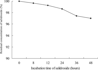
Figure 6 Protective effects of salidroside on hypoxia/reoxygenation injury. Confluent PC12 cells were pretreated with different concentrations of salidroside in 96-well plates for 16 h. Then, the cells were exposed to 10 mM Na2S2O4 for 1 h for hypoxic damage. After hypoxic incubation, the medium was replaced with the former concentration of salidroside for reoxygenation for 2 h. Cell viability was determined by the MTT assay and the data were expressed as X ± SD, n = 3. *p < 0.05, **p < 0.01, and ***p < 0.001 in comparison with hypoxia/reoxygenation injury group cells.
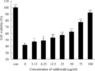
Figure 7 Morphologic inspection of PC12 cells. Confluent PC12 cells were pretreated with different concentrations of salidroside in 96-well plates for 16 h. Then, the cells were exposed to 10 mM Na2S2O4 for 1 h for hypoxic damage. After hypoxic incubation, the medium was replaced with the former concentration of salidroside for reoxygenation for 2 h. Salidroside could prevent the cells from morphologic changes: (1) control group (without salidroside and Na2S2O4), (2) hypoxia/reoxygenation injury group, (3) hypoxia/reoxygenation injury and 100 µg/mL salidroside, (4) hypoxia/reoxygenation injury and 50 µg/mL salidroside, (5) hypoxia/reoxygenation injury and 12.5 µg/mL salidroside.
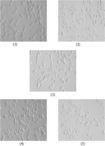
Figure 8 Effects of salidroside on extracelluar LDH activity in PC12 cells. Confluent cells were preincubated with various concentrations of salidroside for 16 h. After hypoxia/reoxygenation injury, the medium was collected and the activity of LDH was determined using LDH test kits according to the manufacturer's protocol. The data were expressed as X ± SD, n = 3. *p < 0.05, **p < 0.01, and ***p < 0.001 in comparison with hypoxia/reoxygenation injury group cells.
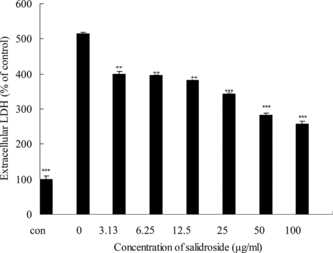
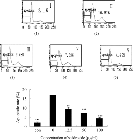
Figure 9 Effects of salidroside on the apoptotic rate in PC12 cells. Apoptotic rate was measured by flow cytometry. (1) control group (without salidroside and Na2S2O4), (2) hypoxia/reoxygenation injury group, (3) hypoxia/reoxygenation injury and 12.5 µg/mL salidroside, (4) hypoxia/reoxygenation injury and 50 µg/mL salidroside, (5) hypoxia/reoxygenation injury and 100 µg/mL salidroside. The data were expressed as X ± SD, n = 3. **p < 0.01 and ***p < 0.001 in comparison with hypoxia/reoxygenation injury group cells.