2,320
Views
13
CrossRef citations to date
0
Altmetric
Research Article

Anti-inflammatory activities of Echinacea extracts do not correlate with traditional marker components

, &
Pages 430-435 | Received 09 Jun 2008, Accepted 09 Sep 2008, Published online: 01 May 2009

Figures & data

Figure 1. Anti-cytokine effects of E. purpurea root extracts. BEAS-2B cells were infected with rhinovirus type 14 (1 pfu/cell) for 60 min at 35°C, followed by 250 μg/mL of the indicated fraction of E. purpurea root extract. Controls received no virus. Solvent controls showed no effect. After 48 h, cell supernatants were removed for assay of IL-6 and IL-8 by standard ELISA tests. Standard curves were constructed for each experiment and the absorbance readings at 450 nm were converted into pg/mL. Composition of marker compounds is shown below the figure.
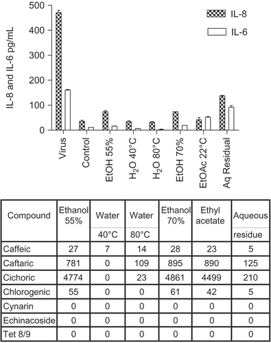
Figure 1.  Anti-cytokine effects of E. purpurea root extracts. BEAS-2B cells were infected with rhinovirus type 14 (1 pfu/cell) for 60 min at 35°C, followed by 250 μg/mL of the indicated fraction of E. purpurea root extract. Controls received no virus. Solvent controls showed no effect. After 48 h, cell supernatants were removed for assay of IL-6 and IL-8 by standard ELISA tests. Standard curves were constructed for each experiment and the absorbance readings at 450 nm were converted into pg/mL. Composition of marker compounds is shown below the figure.

Figure 2. Anti-cytokine effects of E. purpurea leaf plus stem extracts. BEAS-2B cells were infected with rhinovirus type 14 (1 pfu/cell) for 60 min at 35°C, followed by 250 μg/mL of the indicated fraction of E. purpurea extract. Controls received no virus. After 48 h, cell supernatants were removed for assay of IL-6 and IL-8 by standard ELISA tests. Standard curves were constructed for each experiment and the absorbance readings at 450 nm were converted into pg/mL. Not all solvent fractions were available for these tests. Only IL-8 is shown in this graph. Similar results were obtained for IL-6. Composition of marker compounds is shown below the figure.
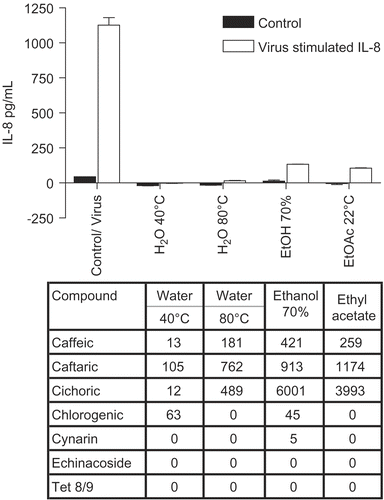
Figure 2.  Anti-cytokine effects of E. purpurea leaf plus stem extracts. BEAS-2B cells were infected with rhinovirus type 14 (1 pfu/cell) for 60 min at 35°C, followed by 250 μg/mL of the indicated fraction of E. purpurea extract. Controls received no virus. After 48 h, cell supernatants were removed for assay of IL-6 and IL-8 by standard ELISA tests. Standard curves were constructed for each experiment and the absorbance readings at 450 nm were converted into pg/mL. Not all solvent fractions were available for these tests. Only IL-8 is shown in this graph. Similar results were obtained for IL-6. Composition of marker compounds is shown below the figure.

Figure 3. Anti-cytokine effects of E. purpurea flower extracts. BEAS-2B cells were infected with rhinovirus type 14 (1 pfu/cell) for 60 min at 35°C, followed by 250 μg/mL of the indicated fraction of E. purpurea flower extract. Controls received no virus. After 48 h, cell supernatants were removed for assay of IL-6 and IL-8 by standard ELISA tests. Standard curves were constructed for each experiment and the absorbance readings at 450 nm were converted into pg/mL. Only IL-8 is shown in this graph; similar results were obtained for IL-6. Composition of marker compounds is shown below the figure.
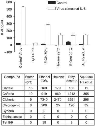
Figure 3.  Anti-cytokine effects of E. purpurea flower extracts. BEAS-2B cells were infected with rhinovirus type 14 (1 pfu/cell) for 60 min at 35°C, followed by 250 μg/mL of the indicated fraction of E. purpurea flower extract. Controls received no virus. After 48 h, cell supernatants were removed for assay of IL-6 and IL-8 by standard ELISA tests. Standard curves were constructed for each experiment and the absorbance readings at 450 nm were converted into pg/mL. Only IL-8 is shown in this graph; similar results were obtained for IL-6. Composition of marker compounds is shown below the figure.

Figure 4. Anti-cytokine effects of polysaccharide-enriched fractions. BEAS-2B cells were infected with rhinovirus type 14 (1 pfu/cell) for 60 min at 35°C, followed by 250 μg/mL of the indicated fraction derived from E. purpurea aqueous pressed juice. Controls received no virus. After 48 h, cell supernatants were removed for assay of IL-6 and IL-8 by standard ELISA tests. Standard curves were constructed for each experiment and the absorbance readings at 450 nm were converted into pg/mL. Polysaccharide-enriched fraction, P50 fraction (polysaccharides > 100 kDa) isolated from pressed juice; AGP fraction, arabinogalactan isolated by SEC 1 column from the polysaccharide fraction.
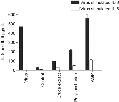
Figure 4.  Anti-cytokine effects of polysaccharide-enriched fractions. BEAS-2B cells were infected with rhinovirus type 14 (1 pfu/cell) for 60 min at 35°C, followed by 250 μg/mL of the indicated fraction derived from E. purpurea aqueous pressed juice. Controls received no virus. After 48 h, cell supernatants were removed for assay of IL-6 and IL-8 by standard ELISA tests. Standard curves were constructed for each experiment and the absorbance readings at 450 nm were converted into pg/mL. Polysaccharide-enriched fraction, P50 fraction (polysaccharides > 100 kDa) isolated from pressed juice; AGP fraction, arabinogalactan isolated by SEC 1 column from the polysaccharide fraction.

Figure 5. Anti-cytokine effects of E. angustifolia root extracts. BEAS-2B cells were infected with rhinovirus type 14 (1 pfu/cell) for 60 min at 35°C, followed by 250 μg/mL of the indicated fraction of E. angustifolia root extract. Controls received no virus. After 48 h, cell supernatants were removed for assay of IL-6 and IL-8 by standard ELISA tests. Standard curves were constructed for each experiment and the absorbance readings at 450 nm were converted into pg/mL. Only IL-8 is shown; the results for IL-6 were similar. Not all solvent fractions were available for this experiment. Composition of marker compounds is shown below the figure.
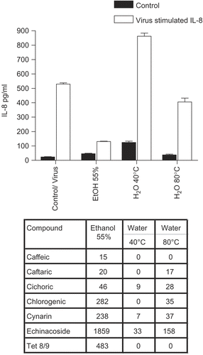
Figure 5.  Anti-cytokine effects of E. angustifolia root extracts. BEAS-2B cells were infected with rhinovirus type 14 (1 pfu/cell) for 60 min at 35°C, followed by 250 μg/mL of the indicated fraction of E. angustifolia root extract. Controls received no virus. After 48 h, cell supernatants were removed for assay of IL-6 and IL-8 by standard ELISA tests. Standard curves were constructed for each experiment and the absorbance readings at 450 nm were converted into pg/mL. Only IL-8 is shown; the results for IL-6 were similar. Not all solvent fractions were available for this experiment. Composition of marker compounds is shown below the figure.

Figure 6. Anti-cytokine effects of E. pallida root extracts. BEAS-2B cells were infected with rhinovirus type 14 (1 pfu/cell) for 60 min at 35°C, followed by 250 μg/mL of the indicated fraction of E. pallida root extract. Controls received no virus. After 48 h, cell supernatants were removed for assay of IL-6 and IL-8 by standard ELISA tests. Standard curves were constructed for each experiment and the absorbance readings at 450 nm were converted into pg/mL. Only IL-8 data are shown; the results for IL-6 were similar. Composition of marker compounds is shown below the figure.
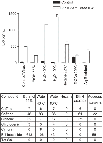
Figure 6.  Anti-cytokine effects of E. pallida root extracts. BEAS-2B cells were infected with rhinovirus type 14 (1 pfu/cell) for 60 min at 35°C, followed by 250 μg/mL of the indicated fraction of E. pallida root extract. Controls received no virus. After 48 h, cell supernatants were removed for assay of IL-6 and IL-8 by standard ELISA tests. Standard curves were constructed for each experiment and the absorbance readings at 450 nm were converted into pg/mL. Only IL-8 data are shown; the results for IL-6 were similar. Composition of marker compounds is shown below the figure.

Reprints and Corporate Permissions

Please note: Selecting permissions does not provide access to the full text of the article, please see our help page How do I view content?

To request a reprint or corporate permissions for this article, please click on the relevant link below:

Academic Permissions

Please note: Selecting permissions does not provide access to the full text of the article, please see our help page How do I view content?

Obtain permissions instantly via Rightslink by clicking on the button below:

If you are unable to obtain permissions via Rightslink, please complete and submit this Permissions form. For more information, please visit our Permissions help page.