2,279
Views
13
CrossRef citations to date
0
Altmetric
Research Article

In vivo and in vitro antiviral activity of five Tibetan medicinal plant extracts against herpes simplex virus type 2 infection

, , , , , , & show all
Pages 598-607 | Received 29 Oct 2008, Accepted 18 Mar 2009, Published online: 22 Jun 2009

Figures & data

Table 1. EC50 and CC50 determinations for the medicinal plant extracts.

Figure 1. Virucidal activity of extracts against HSV-2. 100 PFU HSV-2 was studied on each test extract at various concentration (0.25, 0.5, 1, 2, 4 mg/mL except 0.06, 0.13, 0.25, 0.5, 1 mg/mL for L. brevituba, respectively) for 1 h at 37°C. Each point represents the mean ± SD of three independent experiments.
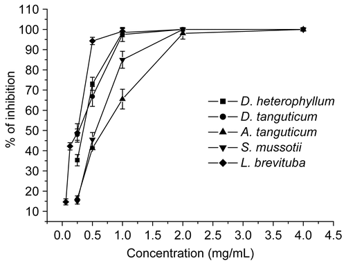
Figure 1.  Virucidal activity of extracts against HSV-2. 100 PFU HSV-2 was studied on each test extract at various concentration (0.25, 0.5, 1, 2, 4 mg/mL except 0.06, 0.13, 0.25, 0.5, 1 mg/mL for L. brevituba, respectively) for 1 h at 37°C. Each point represents the mean ± SD of three independent experiments.

Figure 4. Effect of ACV on HSV-2 infectivity, attachment and biosynthesis to Vero cells. The tested concentration of ACV was 0.03, 0.06, 0.13, 0.25, 0.5 mg/mL. Each point represents the mean ± SD of three independent experiments. A, HSV-2 was mixed with various concentrations of ACV for 1 h at 37°C; B, cells treated with ACV before virus infection; C, cells treated with ACV after virus infection.
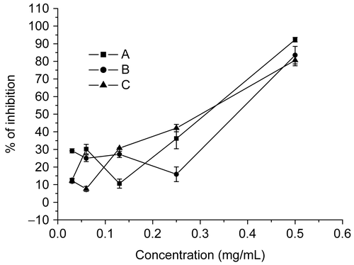
Figure 4.  Effect of ACV on HSV-2 infectivity, attachment and biosynthesis to Vero cells. The tested concentration of ACV was 0.03, 0.06, 0.13, 0.25, 0.5 mg/mL. Each point represents the mean ± SD of three independent experiments. A, HSV-2 was mixed with various concentrations of ACV for 1 h at 37°C; B, cells treated with ACV before virus infection; C, cells treated with ACV after virus infection.

Figure 2. Effect of extracts on HSV-2 attachment to Vero cells. Vero cell monolayer in 24-well plates were treated with extracts before virus infection at various concentration (0.25, 0.5, 1, 2, 4 mg/mL except 0.06, 0.13, 0.25, 0.5, 1 mg/mL for L. brevituba, respectively). Each point represents the mean ± SD of three independent experiments.
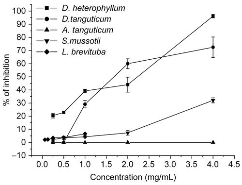
Figure 2.  Effect of extracts on HSV-2 attachment to Vero cells. Vero cell monolayer in 24-well plates were treated with extracts before virus infection at various concentration (0.25, 0.5, 1, 2, 4 mg/mL except 0.06, 0.13, 0.25, 0.5, 1 mg/mL for L. brevituba, respectively). Each point represents the mean ± SD of three independent experiments.
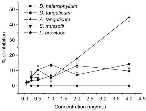
Figure 3. Effect of extracts on HSV-2 biosynthesis to Vero cells. Vero cell monolayer in 24-well plates were treated with extracts after virus infection at various concentration (0.25, 0.5, 1, 2, 4 mg/ml except 0.06, 0.13, 0.25, 0.5, 1 mg/mL for L. brevituba, respectively). Each point represents the mean ± SD of three independent experiments.

Figure 3.  Effect of extracts on HSV-2 biosynthesis to Vero cells. Vero cell monolayer in 24-well plates were treated with extracts after virus infection at various concentration (0.25, 0.5, 1, 2, 4 mg/ml except 0.06, 0.13, 0.25, 0.5, 1 mg/mL for L. brevituba, respectively). Each point represents the mean ± SD of three independent experiments.

Table 2. The effect of extracts against HSV DNA synthesis quantitated using a quantitative real-time PCR.

Table 3. The effect of extracts against HSV-2-induced encephalitis in mice.

Reprints and Corporate Permissions

Please note: Selecting permissions does not provide access to the full text of the article, please see our help page How do I view content?

To request a reprint or corporate permissions for this article, please click on the relevant link below:

Academic Permissions

Please note: Selecting permissions does not provide access to the full text of the article, please see our help page How do I view content?

Obtain permissions instantly via Rightslink by clicking on the button below:

If you are unable to obtain permissions via Rightslink, please complete and submit this Permissions form. For more information, please visit our Permissions help page.